Recurred Adenoid Cystic Carcinoma of Lacrimal Gland with Aggressive Local Invasion to the Maxillary Bone Marrow without Increased Uptake in PET-CT
- Affiliations
-
- 1Department of Ophthalmology, Severance Hospital, Institute of Vision Research, Yonsei University College of Medicine, Seoul, Korea. yoonjs@yuhs.ac
- 2Department of Pathology, Yonsei University College of Medicine, Seoul, Korea.
- KMID: 2360141
- DOI: http://doi.org/10.3341/kjo.2015.29.1.68
Abstract
- No abstract available.
MeSH Terms
-
Bone Marrow/*pathology/radiography/radionuclide imaging
Carcinoma, Adenoid Cystic/*diagnosis
Eye Neoplasms/*diagnosis
Female
Humans
Lacrimal Apparatus/*pathology/radiography/radionuclide imaging
Lacrimal Apparatus Diseases/*diagnosis
Maxilla
Middle Aged
Neoplasm Invasiveness
Neoplasm Recurrence, Local
*Positron-Emission Tomography
*Tomography, X-Ray Computed
Figure
-
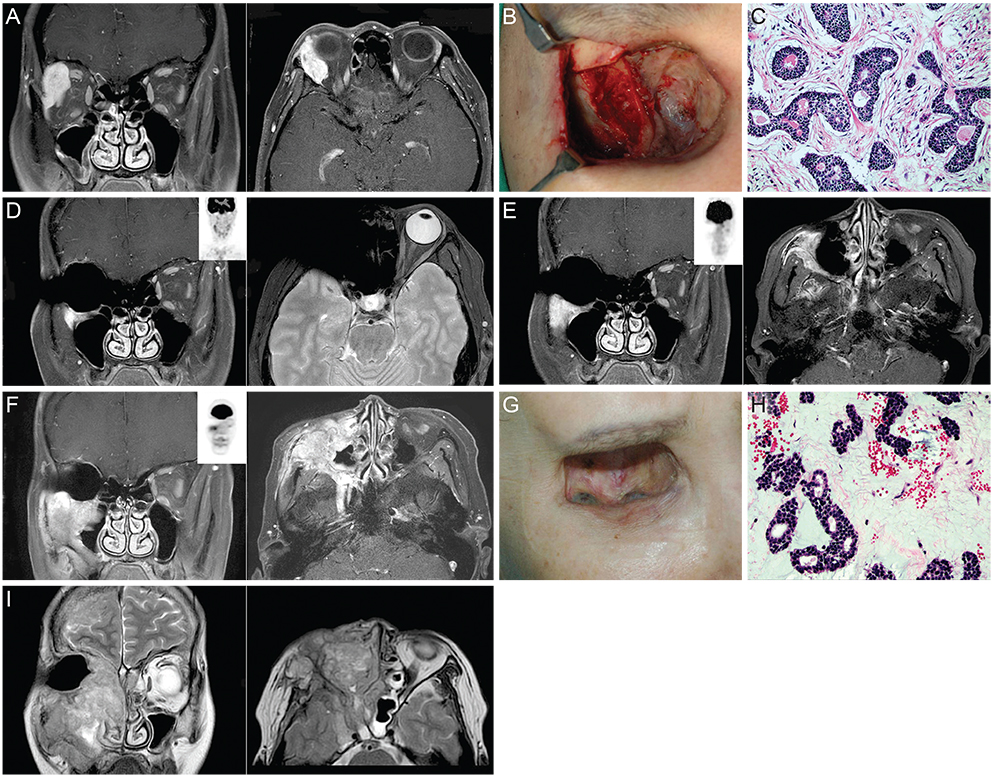
Fig. 1 (A) Coronal and axial views of orbit magnetic resonance imaging (MRI) taken preoperatively, revealing an approximately 3-cm-sized enhancing lesion in the right lacrimal gland with bony invasion. (B) Photograph of the exposed lateral zygoma bone during operation which was pathologically proven to have cancer infiltration. (C) Histologic section of primary cancer of stage T4bN0M0, cribriform type with lymphovascular invasion (H&E, ×100). (D) Coronal and axial views of orbit MRI with positron emission tomography-computed tomography (PET-CT) scan contrast coronal images in the middle taken 1 year after exenteration, showing a focal enhancing lesion in the right anterior maxilla bone without uptake in the PET-CT scan. (E) Two years postoperatively, with increased extent of the enhancing lesion, yet still no visible uptake in the PET-CT scan. (F) Three and half years after local surgery, showing a markedly increased infiltrating mass along the entire maxillary sinus wall with corresponding fludeoxyglucose uptake in the PET-CT scan. (G) Photograph of an elevated pigmented mass along the inferolateral margin of the exenterated orbit 3.5 years after exenteration. (H) Histologic section of recurred cancer (H&E, ×100). The pathologic grade was not different, but the proportion of the mesenchymal component differed in that the primary cancer contained more fibrous components whereas the recurred cancer had more myxoid components. (I) Coronal and axial views of orbit MRI 4.5 years postoperatively, showing the right maxillary sinus mass with intracranial extension, brain edema, and infiltration into the left orbital apex.
Reference
-
1. Esmaeli B, Ahmadi MA, Youssef A, et al. Outcomes in patients with adenoid cystic carcinoma of the lacrimal gland. Ophthal Plast Reconstr Surg. 2004; 20:22–26.2. Tse DT, Kossler AL, Feuer WJ, Benedetto PW. Long-term outcomes of neoadjuvant intra-arterial cytoreductive chemotherapy for lacrimal gland adenoid cystic carcinoma. Ophthalmology. 2013; 120:1313–1323.3. Fellman M, Carter K, Call CB, Esmaeli B. Disease recurrence after intraarterial chemotherapy in 2 patients with adenoid cystic carcinoma of lacrimal gland. Can J Ophthalmol. 2013; 48:e17–e18.4. Lee JY, Choi JY, Ko YH, et al. 18F-FDG PET/CT in patients with initially diagnosed adenoid cystic carcinoma of the head and neck: clinicoplathologic correlation. Nucl Med Mol Imaging. 2009; 43:395–401.5. Qin W, Chong R, Huang X, et al. Adenoid cystic carcinoma of the lacrimal gland: CT and MRI findings. Eur J Ophthalmol. 2012; 22:316–319.
- Full Text Links
-
- Actions
-
Cited
- CITED
-
- Close
- Share
-
- Similar articles
-
- Extensive and aggressive growth of adenoid cysticcarcinoma in the lacrimal gland
- A Case of Adenoid Cystic CArcinoma of Unusually Located Lacrimal Gland
- En Bloc Orbitectomy for Recurred Adenoid Cystic Carcinoma of the Lacrimal Gland
- Lacrimal gland adenoid cystic carcinoma: report of an unusual case with literature review
- Skin Metastasis of Adenoid Cystic Carcinoma of Parotid Gland
