J Korean Ophthalmol Soc.
2016 Nov;57(11):1786-1789. 10.3341/jkos.2016.57.11.1786.
A Case of Primary Conjunctival Giant Cell Tumor
- Affiliations
-
- 1Department of Ophthalmology, Busan Paik Hospital, Inje University College of Medicine, Busan, Korea. Sixqueen51@naver.com
- KMID: 2357737
- DOI: http://doi.org/10.3341/jkos.2016.57.11.1786
Abstract
- PURPOSE
To report a case of primary conjunctival giant cell tumor (GCT).
CASE SUMMARY
A 67-year-old female visited our clinic with the chief complaint of a 10-year-history of conjunctival mass in the left eye. The patient had no marked changes in the mass size, and her visual acuity and intraocular pressure were within the normal range. The protruding conjunctival mass invaded the limbal area at the 8 o'clock direction in the left eye and was 5 × 4 × 2 mm in size. Moreover, the pink-colored mass had a lobulated shape with a well-defined margin. In the adjacent mass region, concurrent presence of the conjunctival injection was observed. However, the patient did not exhibit pain or tenderness. We performed wide excision of the conjunctival mass concomitantly with amniotic membrane transplantation. Then, histopathological examinations and immunohistochemical staining of the surgical site were performed. On histopathology, the patient had findings suggestive of GCT. Additionally, immunohistochemistry was positive for CD68 and vimentin. leading to the final diagnosis of GCT.
CONCLUSIONS
To our knowledge, this is the first case of GCT of the conjunctiva, which has not been described in the literature. Our case highlights the importance of differential diagnosis from other protuberant conjunctival tumors. A complete removal of GCT of the conjunctiva and a recovery of aesthetic outcomes can be achieved by surgical excision of the mass.
MeSH Terms
Figure
-

Figure 1. Photograph of the conjunctival mass at medial side of left eye. Well circumscribed, lobulated, protruding pinkish mass.
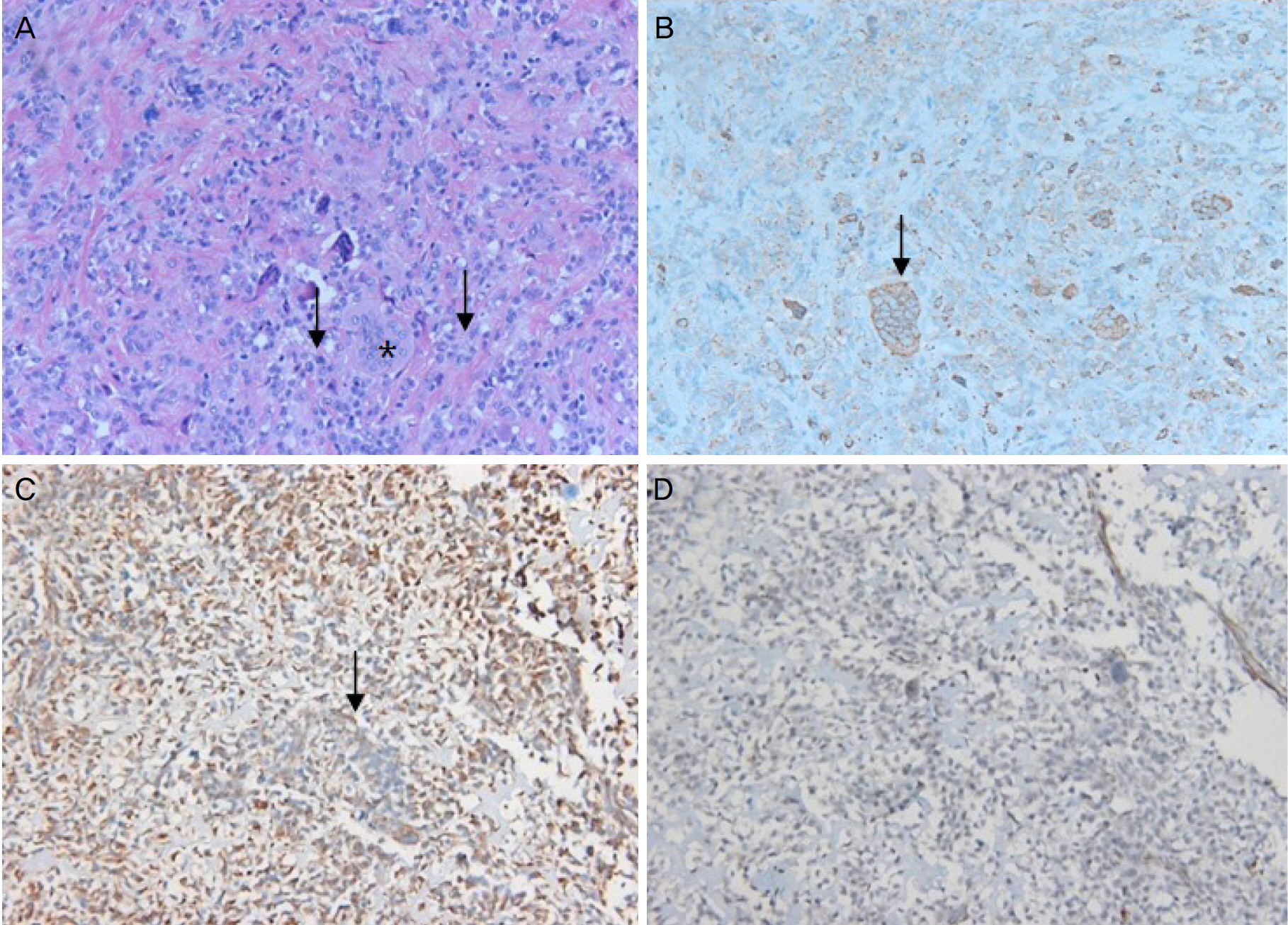
Figure 2. Histological and Immunohistochemical examination of conjunctival giant cell tumor. (A) Multinucleated giant cells (*) interspersed among mononuclear cells (arrows) (H&E ×200). (B) Positive staining for CD68 (arrow) (×200). (C) Positive staining for Vimentin (arrow) (×200). (D) Negative staining for smooth muscle actin (×200).
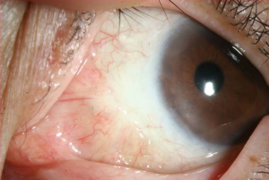
Figure 3. Conjunctival appearance at 6months after surgical resection. The mass was completely removed and there was no local recurrence.
Reference
-
References
1. Plowman RS, Nguyen BD. Primary pulmonary giant cell tumor: (18)F-FDG PET/CT imaging. Rev Esp Med Nucl Imagen Mol. 2016; 35:274–6.2. Li C, Zheng X, Ghert M, et al. Expressions and clinical abdominal of factors related to giant cell tumor of bone. Int J Clin Exp Med. 2015; 8:22509–14. eCollection 2015.3. Sobti A, Agrawal P, Agarwala S, Agarwal M. Giant cell tumor of bone – an overview. Arch Bone Jt Surg. 2016; 4:2–9.4. Yamagishi T, Kawashima H, Ogose A, et al. Disappearance of giant cells and presence of newly formed bone in the pulmonary metastasis of a sacral giant-cell tumor following denosumab abdominal: a case report. Oncol Lett. 2016; 11:243–6.5. Yu X, Guo R, Fan C, et al. Aneurysmal bone cyst secondary to a giant cell tumor of the patella: a case report. Oncol Lett. 2016; 11:1481–5.
Article6. Skubitz KM. Giant cell tumor of bone: current treatment options. Curr Treat Options Oncol. 2014; 15:507–18.
Article7. López-Pousa A, Martín Broto J, Garrido T, Vázquez J. Giant cell tumour of bone: new treatments in development. Clin Transl Oncol. 2015; 17:419–30.
Article8. Wang DD, Zheng YM, Teng LH, et al. Benign giant-cell tumor of the common bile duct: a case report. World J Gastroenterol. 2014; 20:15448–53.
Article9. Temesgen WM, Wachtel M, Dissanaike S. Osteoclastic giant cell tumor of the pancreas. Int J Surg Case Rep. 2014; 5:175–9.
Article10. Manidakis N, Polyzois I, Tsialogiannis E, et al. Metastatic abdominal melanoma of the conjunctiva: a case report. Cases J. 2009; 2:125.11. López-Meca IC, Alcaraz-Mateos E, Córdoba-Polo C, BelmonteMartínez J. Atypical melanocytic nevus of the limbus. A case report. Arch Soc Esp Oftalmol. 2014; 89:265–8.
Article12. Pfister H, Fuchs PG, Völcker HE. Human papillomavirus DNA in conjunctival papilloma. Graefes Arch Clin Exp Ophthalmol. 1985; 223:164–7.
Article13. Buggage RR, Smith JA, Shen D, Chan CC. Conjunctival papil-lomas caused by human papillomavirus type 33. Arch Ophthalmol. 2002; 120:202–4.
Article
