Conservative approach to recurrent calcifying cystic odontogenic tumor occupying the maxillary sinus: a case report
- Affiliations
-
- 1Department of Oral and Maxillofacial Surgery, School of Dentistry, Chonbuk National University, Jeonju, Korea. omfskso@jbnu.ac.kr
- 2Research Institute of Clinical Medicine of Chonbuk National University-Biomedical Research Institute of Chonbuk National University Hospital, Jeonju, Korea.
- KMID: 2356260
- DOI: http://doi.org/10.5125/jkaoms.2016.42.5.315
Abstract
- Calcifying cystic odontogenic tumor (CCOT) is an uncommon benign cystic neoplasm of the jaw that develops from the odontogenic epithelium. Invasion into the maxillary sinus by a CCOT is not a typical, and the recurrence of the cystic variant of CCOT in the posterior maxilla is rare. This report describes a recurrent CCOT occupying most of the maxillary sinus of a 24-year-old male patient. As a treatment, marsupialization was carried out as a means of decompression, and the involved teeth were all endodontically treated. Afterward, surgical enucleation was performed. The size of the lesion continued to shrink after marsupialization, and the maxillary sinus restored its volume. This patient has been followed-up for 3 years after the surgery, and there have not been any signs of recurrence.
MeSH Terms
Figure
-
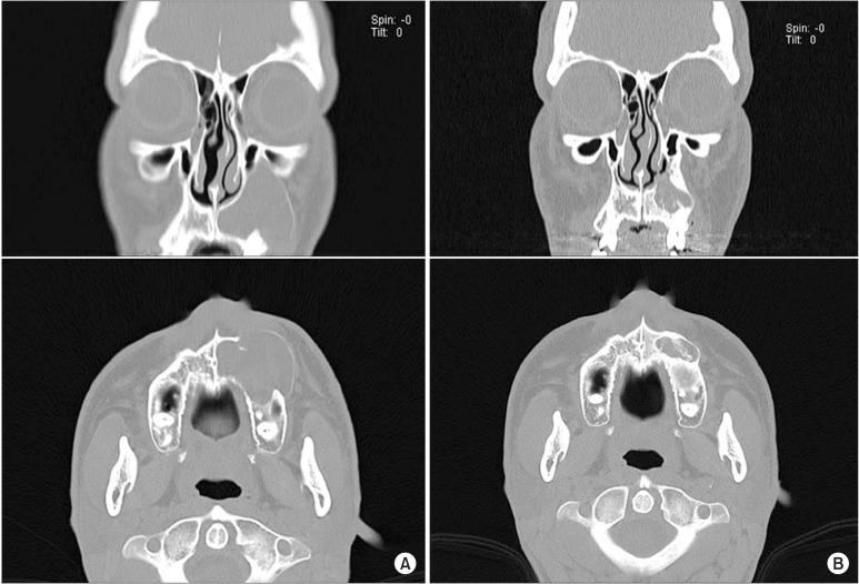
Fig. 1 Computed tomography shows recurrence of the lesion after the first functional endoscopic sinus surgery (FESS) procedure. A. Expansile cyst-like lesion at the first visit. B. A recurrent lesion two years after the first FESS.
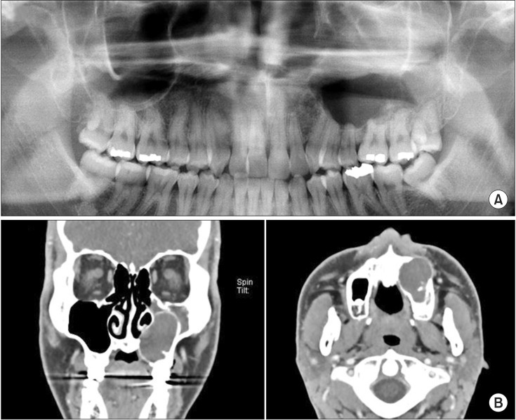
Fig. 2 When the patient was referred to our department, three years after the second functional endoscopic sinus surgery procedure, he showed recurrence in the same region. A. A panoramic radiograph revealing the large extension of unilocular radiolucency in the left maxilla. B. Computed tomography scan showing a large lesion in the left maxillary sinus expanding up to the inferior orbital floor and projecting into the nasal cavity.
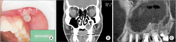
Fig. 3 A. Cyst marsupialization and placement of an acrylic tube for drainage. B. Five months computed tomography (CT) image after the endodontic treatment and marsupialization. C. Cone-beam CT scan showing the regressed lesion and new bone deposition around the lesion.

Fig. 4 Surgical removal of the lesion through enucleation.
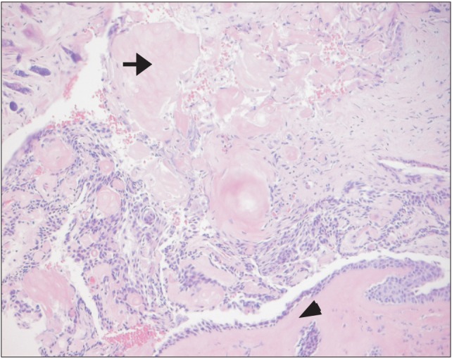
Fig. 5 Histopathologic examination revealing the presence of several ghost cells within the cyst lining and dystrophic calcification (arrow, ghost cell; arrow head, calcifying dentinoid material; H&E staining, ×200).

Fig. 6 Postoperative panoramic view and computed tomography showing the reduced size of the lesion without any recurrences. A. Six months postoperative. B. Thirty-six months postoperative.
Reference
-
1. Gorlin RJ, Pindborg JJ, Clausen FP, Vickers RA. The calcifying odontogenic cyst: a possible analogue of the cutaneous calcifying epithelioma of Malherbe. Oral Surg Oral Med Oral Pathol. 1962; 15:1235–1243. PMID: 13949298.2. Praetorius F, Hjørting-Hansen E, Gorlin RJ, Vickers RA. Calcifying odontogenic cyst. Range, variations and neoplastic potential. Acta Odontol Scand. 1981; 39:227–240. PMID: 6948493.3. Neville BW, Damm DD, Allen CM, Bouquot J. Oral and maxillofacial pathology. 3rd ed. St. Louis: Saunders;2008. p. 695–697.4. Reyes D, Villanueva J, Espinosa S, Cornejo M. Odontogenic calcificant cystic tumor: a report of two clinical cases. Med Oral Patol Oral Cir Bucal. 2007; 12:E126–E129. PMID: 17322800.5. Thinakaran M, Sivakumar P, Ramalingam S, Jeddy N, Balaguhan S. Calcifying ghost cell odontogenic cyst: a review on terminologies and classifications. J Oral Maxillofac Pathol. 2012; 16:450–453. PMID: 23248487.
Article6. Buchner A. The central (intraosseous) calcifying odontogenic cyst: an analysis of 215 cases. J Oral Maxillofac Surg. 1991; 49:330–339. PMID: 2005490.
Article7. Iida S, Fukuda Y, Ueda T, Aikawa T, Arizpe JE, Okura M. Calcifying odontogenic cyst: radiologic findings in 11 cases. Oral Surg Oral Med Oral Pathol Oral Radiol Endod. 2006; 101:356–362. PMID: 16504870.
Article8. Chindasombatjaroen J, Poomsawat S, Boonsiriseth K. Two unique cases of calcifying cystic odontogenic tumor in the maxillary posterior region. Oral Surg Oral Med Oral Pathol Oral Radiol. 2014; 118:497–504. PMID: 25201118.
Article9. Hirshberg A, Kaplan I, Buchner A. Calcifying odontogenic cyst associated with odontoma: a possible separate entity (odontocalcifying odontogenic cyst). J Oral Maxillofac Surg. 1994; 52:555–558. PMID: 8189290.
Article10. Chindasombatjaroen J, Poomsawat S, Klongnoi B. Calcifying cystic odontogenic tumor associated with other lesions: case report with cone-beam computed tomography findings. Oral Surg Oral Med Oral Pathol Oral Radiol. 2012; 113:414–420. PMID: 22669147.
Article11. Daniels JS. Recurrent calcifying odontogenic cyst involving the maxillary sinus. Oral Surg Oral Med Oral Pathol Oral Radiol Endod. 2004; 98:660–664. PMID: 15583537.
Article12. Sciubba JJ, Fantasia JE, Kahn LB. Tumors and cysts of the jaws. Washington: Armed Forces Institute of Pathology;2001. p. 43–45.13. Cheng YS, Wright JM, Walstad WR, Finn MD. Calcifying epithelial odontogenic tumor showing microscopic features of potential malignant behavior. Oral Surg Oral Med Oral Pathol Oral Radiol Endod. 2002; 93:287–295. PMID: 11925538.
Article14. Souza LN, Souza AC, Gomes CC, Loyola AM, Durighetto AF Jr, Gomez RS, et al. Conservative treatment of calcifying odontogenic cyst: report of 3 cases. J Oral Maxillofac Surg. 2007; 65:2353–2356. PMID: 17954339.
Article15. Tanimoto K, Tomita S, Aoyama M, Furuki Y, Fujita M, Wada T. Radiographic characteristics of the calcifying odontogenic cyst. Int J Oral Maxillofac Surg. 1988; 17:29–32. PMID: 3127486.
Article16. Struthers P, Shear M. Root resorption by ameloblastomas and cysts of the jaws. Int J Oral Surg. 1976; 5:128–132. PMID: 820661.
Article17. Ledesma-Montes C, Gorlin RJ, Shear M, Prae Torius F, Mosqueda-Taylor A, Altini M, et al. International collaborative study on ghost cell odontogenic tumours: calcifying cystic odontogenic tumour, dentinogenic ghost cell tumour and ghost cell odontogenic carcinoma. J Oral Pathol Med. 2008; 37:302–308. PMID: 18221328.
Article18. Ellis E. Surgical management of oral pathologic lesions. In : Patterson LJ, Ellis E, Hupp JR, Tucker MR, editors. Contemporary oral and maxillofacial surgery. 2nd ed. St. Louis: Mosby-Year Book;1993. p. 526–553.19. Brøndum N, Jensen VJ. Recurrence of keratocysts and decompression treatment. A long-term follow-up of forty-four cases. Oral Surg Oral Med Oral Pathol. 1991; 72:265–269. PMID: 1717918.20. Schuster D, Cure J, Woodworth BA. Transnasal endoscopic resection of a calcifying cystic odontogenic tumor. Ear Nose Throat J. 2014; 93:E28–E30.
Article
- Full Text Links
-
- Actions
-
Cited
- CITED
-
- Close
- Share
-
- Similar articles
-
- Ghost Cell Odontogenic Carcinoma Arising from Calcifying Cystic Odontogenic Tumor: A Case Report
- Calcifying Odontogenic Cyst Associated with Maxillary Sinus: Case Report
- Case Report of Compound Odontoma Extending Into Maxillary Sinus
- Extensive Adenomatoid Odontogenic Tumor of the Maxilla: A Case Report of Conservative Surgical Excision and Orthodontic Alignment of Impacted Canine
- Dentinogenic Ghost Cell Tumor: A Case Report and Review of Literature
