Polycyclic Annular Lesion Masquerading as Lupus Erythematosus and Emerging as Tinea Faciei Incognito
- Affiliations
-
- 1Department of Dermatology, Korea University College of Medicine, Seoul, Korea. grace79@korea.ac.kr
- KMID: 2352506
- DOI: http://doi.org/10.5021/ad.2015.27.3.322
Abstract
- Tinea incognito is a dermatophytic infection induced by immunosuppressive agents that lacks the classic features of a typical fungal infection. Although the treatment of tinea incognito is simple and relatively easy, its clinical manifestation varies and can masquerade as various skin disorders, causing misdiagnosis and thus preventing prompt and appropriate treatment. Here, we report an interesting case of tinea incognito occurring after topical steroid administration in an immunosuppressed patient with dermatitis artefacta. A 40-year-old female patient who had been taking systemic glucocorticoid for 4 years for chronic inflammatory demyelinating polyneuropathy presented with itching multiple erythematous erosive lesions on the face and upper chest for 2 months. Initial biopsy produced nonspecific findings. The skin lesion was aggravated and became polycyclic and erythematous; after azathioprine was added, her chronic inflammatory demyelinating polyneuropathy became aggravated. A second biopsy confirmed hyphae in the cornified layer. Complete remission was achieved after admonishing oral terbinafine and topical amorolfine.
MeSH Terms
Figure
-

Fig. 1 Clinical photographs of polycyclic annular lesion of tinea faciei incognito. (A) The patient presented with multiple erythematous erosive macules at the initial visit, which was diagnosed as dermatitis artefacta. (B) The skin lesions initially improved but were abruptly aggravated after topical steroid administration for 2 months, showing polycyclic features with fine scales. (C) Enlarged view of polycyclic skin lesions with fine scales. (D) Dramatic remission was observed after 1 week of treatment with systemic and topical anti-fungal agents.
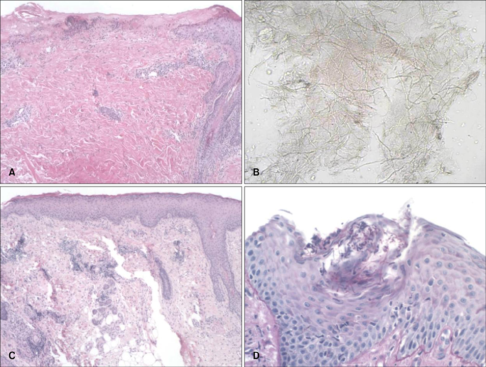
Fig. 2 Histopathological findings of the lesion. (A) The histopathologic examination of initial erosive lesion yielded no specific findings, which led to a diagnosis of dermatitis artefacta (H&E, ×40). (B) The KOH test performed at the margin of a polycyclic scaly skin lesion revealed fugal hyphae (×100). (C) The histopathology of a polycyclic skin lesion on the forehead revealed no evidence of lupus erythematosus (H&E, ×40). (D) Periodic acid-Schiff stain showing fungal hyphae in the cornified layer (×200).
Cited by 1 articles
-
Tinea Incognito with Folliculitis-Like Presentation: A Case Series
Min-Woo Kim, Hyun-Sun Park, Jeong Mo Bae, Hyun-Sun Yoon, Soyun Cho
Ann Dermatol. 2018;30(1):97-99. doi: 10.5021/ad.2018.30.1.97.
Reference
-
1. Romano C, Maritati E, Gianni C. Tinea incognito in Italy: a 15-year survey. Mycoses. 2006; 49:383–387.
Article2. Guenova E, Hoetzenecker W, Schaller M, Röcken M, Fierlbeck G. Tinea incognito hidden under apparently treatment-resistant pemphigus foliaceus. Acta Derm Venereol. 2008; 88:276–277.3. Agostini G, Knöpfel B, Difonzo EM. Universal dermatophytosis (tinea incognito) caused by Trichophyton rubrum. Hautarzt. 1995; 46:190–193.4. Feder HM Jr. Tinea incognito misdiagnosed as erythema migrans. N Engl J Med. 2000; 343:69.
Article5. Jacobs JA, Kolbach DN, Vermeulen AH, Smeets MH, Neuman HA. Tinea incognito due to Trichophytom rubrum after local steroid therapy. Clin Infect Dis. 2001; 33:E142–E144.6. Stein DH. Tineas--superficial dermatophyte infections. Pediatr Rev. 1998; 19:368–372.
Article7. Al-Amiri A, Chatrath V, Bhawan J, Stefanato CM. The periodic acid-Schiff stain in diagnosing tinea: should it be used routinely in inflammatory skin diseases? J Cutan Pathol. 2003; 30:611–615.
Article8. Park YW, Kim DY, Yoon SY, Park GY, Park HS, Yoon HS, et al. 'Clues' for the histological diagnosis of tinea: how reliable are they? Ann Dermatol. 2014; 26:286–288.
Article
- Full Text Links
-
- Actions
-
Cited
- CITED
-
- Close
- Share
-
- Similar articles
-
- A Clinical and Etiological Analysis of Tinea Incognito Over 10 Years: A Single-Center Experience
- Tinea Pseudoimbricata caused by Trichophyton rubrum
- A Case of Tinea Incognito on the Face due to Trichophyton mentagrophytes
- A Case of Subacute Cutaneous Lupus Erythematosus That Simultaneously Presented with Papulosquamous and Annular Lesions
- Tinea Incognito Caused by Application of 0.03% Tacrolimus (Protopic(R)) Ointment in Atopic Dermatitis Patient
