A Case of Acute Angle Closure Caused by Dislocation of Accommodative Intraocular Lens
- Affiliations
-
- 1Myung-Gok Eye Research Institute, Department of Ophthalmology, Kim's Eye Hospital, Konyang University College of Medicine, Seoul, Korea. brainh@hanmail.net
- 2Seoul Ire Eye Clinic, Seoul, Korea.
- KMID: 2351885
- DOI: http://doi.org/10.3341/jkos.2016.57.9.1493
Abstract
- PURPOSE
To report a case of acute angle closure after cataract surgery using an accommodative intraocular lens (IOL), WIOL-CF® (GELMED, Praha, Czech).
CASE SUMMARY
A 46-year-old male patient underwent phacoemulsification and implantation of WIOL-CF® into the capsular bag. Seven months after the surgery, a sudden increase in intraocular pressure (IOP) associated with angle closure was observed. Ultrabiomicroscopy revealed a dislocated WIOL-CF® that was pushing the peripheral iris anteriorly. Despite the use of IOP-lowering medication and peripheral laser iridotomy, IOP was not controlled. After the use of cycloplegics, the angle was widened and IOP decreased; however, after nine days, the WIOL-CF® was completely dislocated into the anterior chamber and so was removed.
CONCLUSIONS
When performing cataract surgery using WIOL-CF®, a possibility of dislocation of IOL and subsequent angle closure should be considered.
MeSH Terms
Figure
-
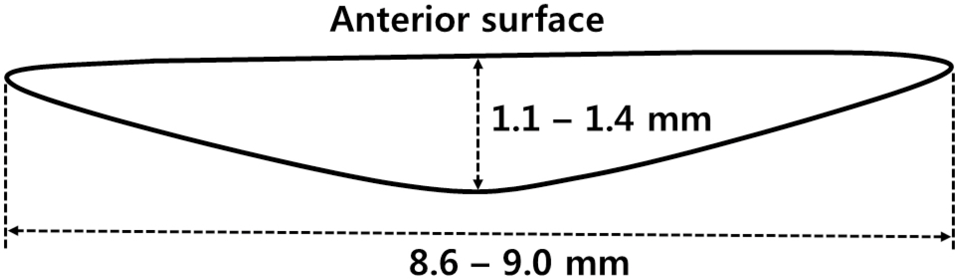
Figure 1. Profile of accommodative intraocular lens, WIOL-CF®(GELMED, Praha, Czech).
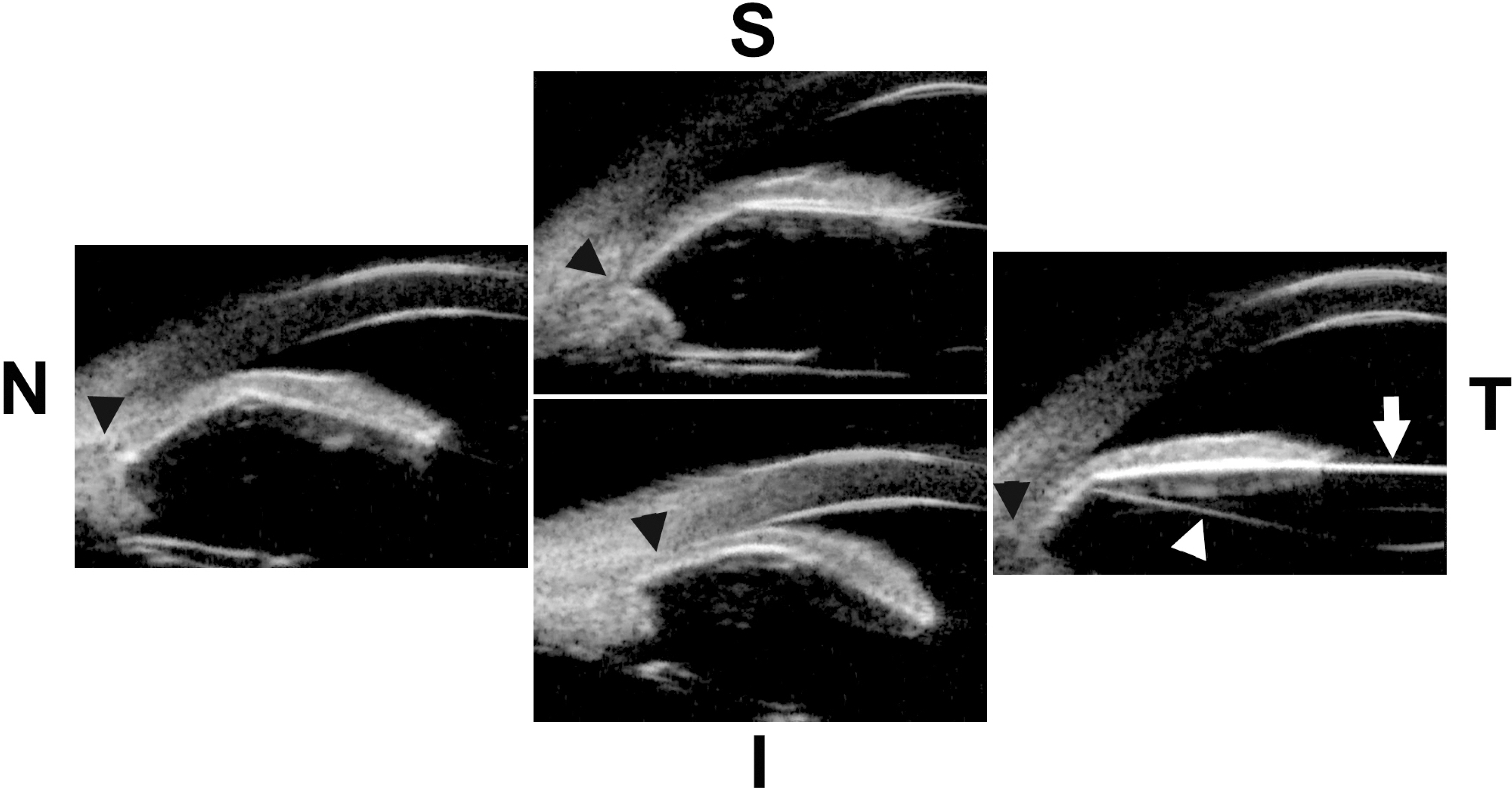
Figure 2. Ultrasound biomicroscopic findings of superior (S), nasal (N), inferior (I), and temporal (T) angle at the time of acute angle closure. White arrow indicates anterior surface of intraocular lens (IOL), white arrowhead indicates posterior surface of IOL, and black arrowheads indicate anterior angle. IOL was dislocated from capsular bag and dislocated IOL was pushing peripheral iris anteriorly. Anterior angles were closed in all quadrants due to anterior displacement of IOL.

Figure 3. Ultrasound biomicroscopic findings of superior (S), nasal (N), inferior (I), and temporal (T) angle after the instillation of cycloplegics. White arrow indicates anterior surface of intraocular lens (IOL), white arrowhead indicates posterior surface of IOL, and black arrowheads indicate anterior angle. IOL moved posteriorly and anterior angle was widened compared to the previous state.

Figure 4. Slit-lamp photograph 9 days after the instillation of cycloplegics. Intraocular lens was completely dislocated into the anterior chamber. White arrows indicate margin of intraocular lens.
Reference
-
References
1. Studeny P, Krizova D, Urminsky J. Clinical experience with the WIOL-CF accommodative bioanalogic intraocular lens: Czech national observational registry. Eur J Ophthalmol. 2016; 26:230–5.
Article2. Pallikaris IG, Portaliou DM, Kymionis GD, et al. Outcomes after accommodative bioanalogic intraocular lens implantation. J Refract Surg. 2014; 30:402–6.
Article3. Lee HS, Park SH, Kim MS. Clinical results and some problems of multifocal apodized diffractive intraocular lens implantation. J Korean Ophthalmol Soc. 2008; 49:1235–41.
Article4. Cheon MH, Lee JE, Kim JH, et al. One-year outcome of monocular implant of aspheric multifocal IOL. J Korean Ophthalmol Soc. 2010; 51:822–28.
Article5. Kang KT, Kim YC. Dislocation of polyfocal full-optics abdominal intraocular lens after neodymium-doped yttrium aluminum garnet capsulotomy in vitrectomized eye. Indian J Ophthalmol. 2013; 61:678–80.
- Full Text Links
-
- Actions
-
Cited
- CITED
-
- Close
- Share
-
- Similar articles
-
- Biometric Measurements in Acute Angle Closure Glaucoma
- Biometric Comparisons between Acute and Chronic Angle Closure Glaucoma
- A Case of Iridoshisis with Angle Closure Glaucoma
- Influence of Lens Factor and Effect of Selected Cataract Extraction on Acute Angle-Closure Glaucoma
- Intraocular Lens Scleral Fixation by Flanged Fixation Using 5-0 Polyprophylene
