Reliability of a New Non-invasive Tear Film Break-up Time Measurement Using a Keratograph
- Affiliations
-
- 1Department of Ophthalmology, Yeungnam University College of Medicine, Daegu, Korea. sbummlee@ynu.ac.kr
- KMID: 2351864
- DOI: http://doi.org/10.3341/jkos.2016.57.9.1354
Abstract
- PURPOSE
To evaluate the repeatability of non-invasive tear film break-up time and identify its relationships with dry eye parameters.
METHODS
A total of 100 participants (50 with dry eye, and 50 in the control group) were enrolled prospectively. Non-invasive keratograph first (NIKf-BUT) and average (NIKav-BUT) break-up times were evaluated 2 times using Keratograph 4 (Oculus, Wetzler, Germany), and then tear film break-up time with fluorescein (FBUT) was measured. The correlation analyses were performed between non-invasive parameters (NIKf-BUT and NIKav-BUT) and FBUT. Intra-observer agreements of NIKf-BUT and NIKav-BUT were assessed using intraclass correlation coefficients (ICC). The receiver operating characteristic (ROC) curve technique was used to evaluate the non-invasive method in the diagnosis of dry eye.
RESULTS
The correlation analyses revealed positive correlation between NIKav-BUT and FBUT in both groups (dry eye; r = 0.66, p < 0.001 and control group; r = 0.77, p < 0.001). The ICCs of NIKf-BUT and NIKav-BUT were 0.72 and 0.94 in the dry eye, respectively, and 0.70 and 0.91 in the control group. NIKav-BUT was not different from FBUT in either group. The areas under the ROC curves of NIKf-BUT and NIKav-BUT were 0.917 and 0.980, respectively.
CONCLUSIONS
The high ICCs verified the repeatability of NIKf-BUT and NIKav-BUT. NIKav-BUT showed no difference from FBUT and positive correlation with FBUT. NIK-BUT showed high diagnostic power and can be considered a new parameter to evaluate dry eye syndrome.
MeSH Terms
Figure
-
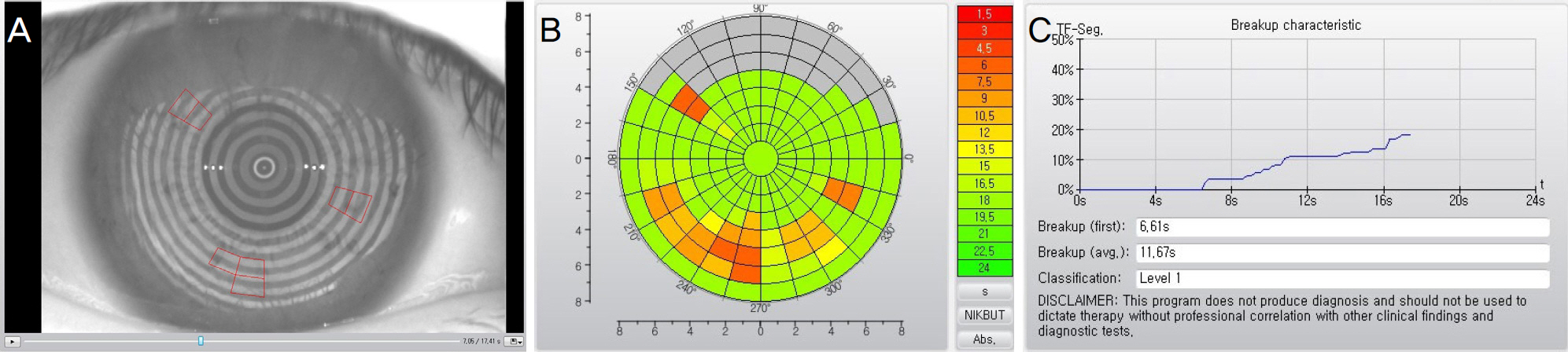
Figure 1. Representative image of non-invasive keratograph tear film break-up time (NIK-BUT) using Oculus Keratograph 4 (Oculus, Wetzler, Germany). (A) A real time image recorded the entire course of break up process. Placido rings were reflected from surface of cornea and their distortions were recorded as the red-framed rectangular break-up units. (B) The final report was summarized as tear film break-up colour-code map. (C) Noninvasive keratograph first break-up time (NIKf-BUT), non-invasive keratograph average break-up time (NIKav-BUT) and break-up progress of break-up units were provided.
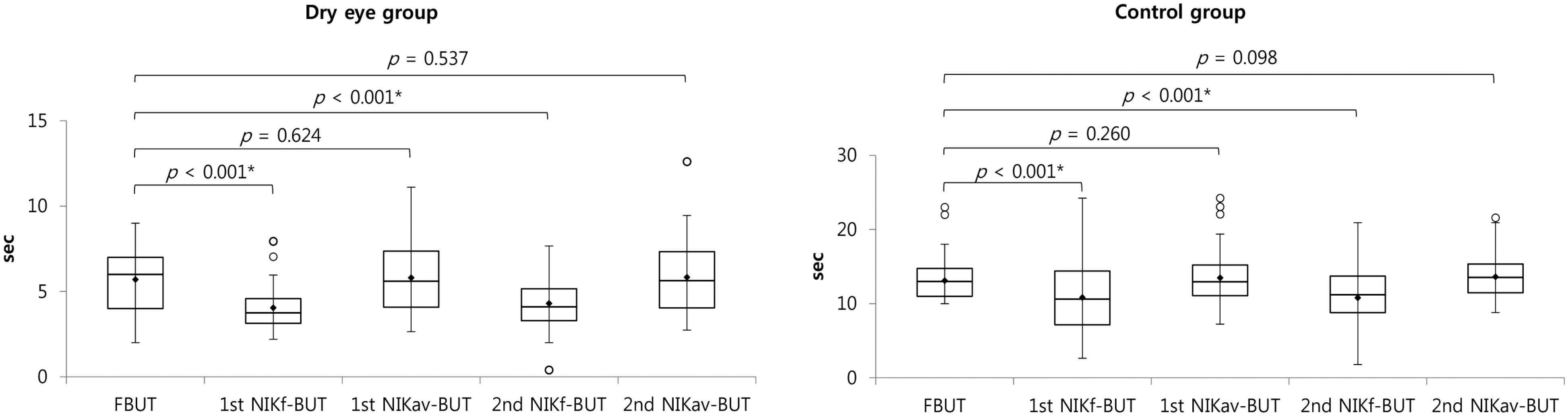
Figure 2. Distribution of non-invasive keratograph tear film break-up time (NIK-BUT) and tear film break-up time with fluorecein (FBUT) using box and whisker plot in the dry eye and control groups. The boxes include 50% of the measured values between 1st and 3rd quartiles and the median (horizontal line). The upper and lower fences indicate 1.5 times the interquartile range (IQR) from 3rd and 1st quartiles. The outliers which are more than 1.5 IQR from the box are shown as circles. The mean of each parameter are shown as diamond. Comparison between FBUT and NIK-BUTs was performed by paired sample t-test. NIKf-BUT = non-invasive keratograph first break-up time; NIKav-BUT = non-invasive keratograph average break-up time.* p-value < 0.05 by paired sample t-test.

Figure 3. Receiver operating characteristic curve (ROC) of non-invasive keratograph tear film break-up time (NIK-BUT). The area under the curve (AUC) is 0.917 in 1st non-invasive keratograph first break-up time (NIKf-BUT) and 0.980 in 1st non-invasive keratograph average break-up time (NIKav-BUT). The cutoff value derived from ROC curve was provided. The difference of AUC between NIKf-BUT and NIKav-BUT was 0.063 and was significant (p = 0.032).
Cited by 1 articles
-
The Use of Keratography to Study Changes on the Ocular Surface after Absorbable Plug Insertion
Hee Jong Shin, Chang-Hyun Park, Kyung Sun Na, Hyun Seung Kim
J Korean Ophthalmol Soc. 2018;59(1):17-22. doi: 10.3341/jkos.2018.59.1.17.
Reference
-
References
1. The definition and classification of dry eye disease: report of the Definition and Classification Subcommittee of the International Dry Eye WorkShop (2007). Ocul Surf. 2007; 5:75–92.2. Hyon JY, Kim HM, Lee D, et al. Korean guidelines for the abdominal and management of dry eye: development and validation of clinical efficacy. Korean J Ophthalmol. 2014; 28:197–206.3. Norn MS. Desiccation of the precorneal film. I. Corneal wet-ting-time. Acta Ophthalmol (Copenh). 1969; 47:865–80.4. Mengher LS, Bron AJ, Tonge SR, Gilbert DJ. Effect of fluorescein instillation on the precorneal tear film stability. Curr Eye Res. 1985; 4:9–12.
Article5. Patel S, Murray D, McKenzie A, et al. Effects of fluorescein on tear breakup time and on tear thinning time. Am J Optom Physiol Opt. 1985; 62:188–90.
Article6. Mengher LS, Bron AJ, Tonge SR, Gilbert DJ. A non-invasive instrument for clinical assessment of the precorneal tear film stability. Curr Eye Res. 1985; 4:1–7.
Article7. Cox SM, Nichols KK, Nichols JJ. Agreement between automated and traditional measures of tear film breakup. Optom Vis Sci. 2015; 92:e257–63.
Article8. Hong J, Sun X, Wei A, et al. Assessment of tear film stability in dry eye with a newly developed keratograph. Cornea. 2013; 32:716–21.
Article9. Jiang Y, Ye H, Xu J, Lu Y. Noninvasive Keratograph assessment of tear film break-up time and location in patients with age-related cataracts and dry eye syndrome. J Int Med Res. 2014; 42:494–502.
Article10. Schiffman RM, Christianson MD, Jacobsen G, et al. Reliability and validity of the Ocular Surface Disease Index. Arch Ophthalmol. 2000; 118:615–21.
Article11. Korb DR. Survey of preferred tests for diagnosis of the tear film and dry eye. Cornea. 2000; 19:483–6.
Article12. Nichols KK, Mitchell GL, Zadnik K. The repeatability of clinical measurements of dry eye. Cornea. 2004; 23:272–85.
Article13. Lee JH, Kee CW. The significance of tear film break-up time in the diagnosis of dry eye syndrome. Korean J Ophthalmol. 1988; 2:69–71.
Article14. Goto E, Tseng SC. Differentiation of lipid tear deficiency dry eye by kinetic analysis of tear interference images. Arch Ophthalmol. 2003; 121:173–80.
Article15. Guillon JP. Use of the Tearscope Plus and attachments in the abdominal examination of the marginal dry eye contact lens patient. Adv Exp Med Biol. 1998; 438:859–67.16. Best N, Drury L, Wolffsohn JS. Clinical evaluation of the Oculus Keratograph. Cont Lens Anterior Eye. 2012; 35:171–4.
Article17. Cho P, Douthwaite W. The relation between invasive and non-invasive tear break-up time. Optom Vis Sci. 1995; 72:17–22.
Article
- Full Text Links
-
- Actions
-
Cited
- CITED
-
- Close
- Share
-
- Similar articles
-
- Effect of Loose Masks on Tear-film Break-up Time
- A Comparison between Keratograph 5M® and IDRA® in Dry Eye Patients
- A Pilot Study of Changes in Tear Film Short-term Dynamics with Infrared Imaging after Phacoemulsification
- Comparison of Each Eye According to the Order of Noninvasive Keratographic Tear Film Evaluation
- Effects of Non-invasive Keratograph Break-Up Time on the Repeatability of Keratometry Measurements
