Craniopharyngiomas : Radiological Differentiation of Two Types
- Affiliations
-
- 1The Russell H. Morgan Department of Radiology and Radiological Sciences, The Johns Hopkins Medical Institutions, Baltimore, MD, USA. leeinho1974@hanmail.net
- 2Department of Radiology, Chungnam National University Hospital, Chungnam National University School of Medicine, Daejeon, Korea.
- 3Department of Pathology, The Johns Hopkins Medical Institutions, Baltimore, MD, USA.
- 4Genometrics Section, Computational and Statistical Genomics Branch, National Human Genome Research Institute, National Institutes of Health, Baltimore, MD, USA.
- KMID: 2351714
- DOI: http://doi.org/10.3340/jkns.2016.59.5.466
Abstract
OBJECTIVE
To determine imaging features that may separate adamantinomatous and papillary variants of craniopharyngiomas given that tumors with adamantinomatous signature features are associated with higher recurrence rates, morbidity, and mortality. We specifically reviewed calcification on CT, T1 bright signal intensity, and cystic change on T2 weighted images for differentiating these two types.
METHODS
We retrospectively reviewed the MRI and CT studies in 38 consecutive patients with pathologically proven craniopharyngiomas between January 2004 and February 2014 for the presence of calcification on CT scans, bright signal intensity on T1 weighted images, and cystic change on T2 weighted images.
RESULTS
Of the 38 craniopharyngiomas, 30 were adamantinomatous type and 8 were papillary type. On CT scans, calcification was present in 25 of 38 tumors. All calcified tumors were adamantinomatous type. Twenty four of 38 tumors had bright signal intensity on T1 weighted images. Of these 24 tumors, 22 (91.7%) were adamantinomatous and 2 were papillary type. Cystic change on T2 weighted images was noted in 37 of 38 tumors; only 1 tumor with papillary type did not show cystic change.
CONCLUSION
T1 bright signal intensity and calcification on CT scans uniformly favor the adamantinomatous type over papillary type of craniopharyngioma in children. However, these findings are more variable in adults where calcification and T1 bright signal intensity occur in 70.6% and 58.8% respectively of adult adamantinomatous types of craniopharyngiomas.
MeSH Terms
Figure
-

Fig. 1 2-year old boy with craniopharyngioma (adamantinomatous type). A : Sagittal T1 weighted image shows diffuse bright signal intensity (arrow) in the sellar and suprasellar mass. B : Non-contrast axial CT scan shows curvilinear high density (arrowheads) in the peripheral portion of mass, indicating calcification.
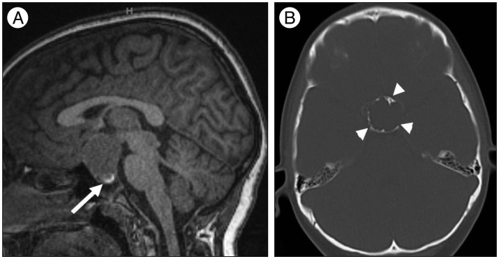
Fig. 2 10-year old boy with craniopharyngioma (adamantinomatous type). A : Sagittal T1 weighted image shows bright signal intensity (arrow) in the lower portion of sellar and suprasellar mass. B : Non-contrast axial CT scan shows curvilinear high density (arrowheads) in the peripheral portion of mass, indicating calcification.

Fig. 3 54-year old woman with craniopharyngioma (papillary type). A : Sagittal T1 weighted image shows no bright signal intensity (arrow) in the sellar and suprasellar mass. B : Non-contrast axial CT scan shows no high density in the mass, indicating calcification.
Reference
-
1. Adamson TE, Wiestler OD, Kleihues P, Yaşargil MG. Correlation of clinical and pathological features in surgically treated craniopharyngiomas. J Neurosurg. 1990; 73:12–17. PMID: 2352012.
Article2. Ahmadi J, Destian S, Apuzzo ML, Segall HD, Zee CS. Cystic fluid in craniopharyngiomas : MR imaging and quantitative analysis. Radiology. 1992; 182:783–785. PMID: 1535894.
Article3. Bunin GR, Surawicz TS, Witman PA, Preston-Martin S, Davis F, Bruner JM. The descriptive epidemiology of craniopharyngioma. J Neurosurg. 1998; 89:547–551. PMID: 9761047.
Article4. Crotty TB, Scheithauer BW, Young WF Jr, Davis DH, Shaw EG, Miller GM, et al. Papillary craniopharyngioma : a clinicopathological study of 48 cases. J Neurosurg. 1995; 83:206–214. PMID: 7616262.5. Curran JG, O'Connor E. Imaging of craniopharyngioma. Childs Nerv Syst. 2005; 21:635–639. PMID: 16078078.
Article6. De Vile CJ, Grant DB, Kendall BE, Neville BG, Stanhope R, Watkins KE. Management of childhood craniopharyngioma : can the morbidity of radical surgery be predicted? J Neurosurg. 1996; 85:73–81. PMID: 8683285.
Article7. Duff J, Meyer FB, Ilstrup DM, Laws ER Jr, Schleck CD, Scheithauer BW. Long-term outcomes for surgically resected craniopharyngiomas. Neurosurgery. 2000; 46:291–302. PMID: 10690718.
Article8. Eldevik OP, Blaivas M, Gabrielsen TO, Hald JK, Chandler WF. Craniopharyngioma : radiologic and histologic findings and recurrence. AJNR Am J Neuroradiol. 1996; 17:1427–2439. PMID: 8883637.9. Fisher PG, Jenab J, Gopldthwaite PT, Tihan T, Wharam MD, Foer DR. Outcomes and failure patterns in childhood craniopharyngiomas. Childs Nerv Syst. 1998; 14:558–563. PMID: 9840379.
Article10. Gautier A, Godbout A, Grosheny C, Tejedor I, Coudert M, Courtillot C, et al. Markers of recurrence and long-term morbidity in craniopharyngioma : a systematic analysis of 171 patients. J Clin Endocrinol Metab. 2012; 97:1258–1267. PMID: 22319039.
Article11. Gupta DK, Ojha BK, Sarkar C, Mahapatra AK, Mehta VS. Recurrence in craniopharyngiomas : analysis of clinical and histological features. J Clin Neurosci. 2006; 13:438–442. PMID: 16678722.
Article12. Jo KW, Shin HJ, Kong DS, Seol HJ, Nam DH, Lee JI. Treatment outcomes of pediatric craniopharyngioma : a 15-year retrospective review of 35 cases. J Korean Neurosurg Soc. 2012; 52:37–41. PMID: 22993676.
Article13. Kim SK, Wang KC, Shin SH, Choe G, Chi JG, Cho BK. Radical excision of pediatric craniopharyngioma : recurrence pattern and prognostic factors. Childs Nerv Syst. 2001; 17:531–536. PMID: 11585327.
Article14. Komotar RJ, Roguski M, Bruce JN. Surgical management of craniopharyngiomas. J Neurooncol. 2009; 92:283–296. PMID: 19357956.
Article15. Miller DC. Pathology of craniopharyngiomas : clinical import of pathological findings. Pediatr Neurosurg. 1994; 21(suppl 1):11–17. PMID: 7841069.
Article16. Mollá E, Martí-Bonmatí L, Revert A, Arana E, Menor F, Dosdá R, et al. Craniopharyngiomas : identification of different semiological patterns with MRI. Eur Radiol. 2002; 12:1829–1836. PMID: 12111075.
Article17. Pekmezci M, Louie J, Gupta N, Bloomer MM, Tihan T. Clinicopathological characteristics of adamantinomatous and papillary craniopharyngiomas : University of California, San Francisco experience 1985-2005. Neurosurgery. 2010; 67:1341–1349. PMID: 20871436.
Article18. Petito CK. Craniopharyngioma : prognostic importance of histologic features. AJNR Am J Neuroradiol. 1996; 17:1441–1442. PMID: 8883638.19. Prabhu VC, Brown HG. The pathogenesis of craniopharyngiomas. Childs Nerv Syst. 2005; 21:622–627. PMID: 15965669.
Article20. Sartoretti-Schefer S, Wichmann W, Aguzzi A, Valavanis A. MR differentiation of adamantinous and squamous-papillary craniopharyngiomas. AJNR Am J Neuroradiol. 1997; 18:77–87. PMID: 9010523.21. Shapiro K, Till K, Grant DN. Craniopharyngiomas in childhood. A rational approach to treatment. J Neurosurg. 1979; 50:617–623. PMID: 430156.22. Szeifert GT, Sipos L, Horváth M, Sarker MH, Major O, Salomváry B, et al. Pathological characteristics of surgically removed craniopharyngiomas : analysis of 131 cases. Acta Neurochir (Wien). 1993; 124:139–143. PMID: 7508161.
Article23. Tavangar SM, Larijani B, Mahta A, Hosseini SM, Mehrazine M, Bandarian F. Craniopharyngioma : a clinicopathological study of 141 cases. Endocr Pathol. 2004; 15:339–344. PMID: 15681858.24. Warakaulle DR, Anslow P. Differential diagnosis of intracranial lesions with high signal on T1 or low signal on T2-weighted MRI. Clin Radiol. 2003; 58:922–933. PMID: 14654024.
Article25. Weiner HL, Wisoff JH, Rosenberg ME, Kupersmith MJ, Cohen H, Zagzag D. Craniopharyngiomas : a clinicopathological analysis of factors predictive of recurrence and functional outcome. Neurosurgery. 1994; 35:1001–1010. PMID: 7885544.26. Zacharia BE, Bruce SS, Goldstein H, Malone HR, Neugut AI, Bruce JN. Incidence, treatment and survival of patients with craniopharyngioma in the surveillance, epidemiology and end results program. Neuro Oncol. 2012; 14:1070–1078. PMID: 22735773.
Article27. Zhang YQ, Wang CC, Ma ZY. Pediatric craniopharyngiomas : clinicomorphological study of 189 cases. Pediatr Neurosurg. 2002; 36:80–84. PMID: 11893889.
Article
- Full Text Links
-
- Actions
-
Cited
- CITED
-
- Close
- Share
-
- Similar articles
-
- Gamma Knife Radiosurgery for Remnant or Recurred Craniopharyngiomas
- MR Differentiation of Craniopharyngioma from Pituitary Macroadenoma
- Should Adjuvant Radiotherapy Be Recommended for Pediatric Craniopharyngiomas?
- Correlation of Treatment Outcome, Histologic Type, and PCNA Labelling Index in Craniopharyngiomas
- The radiological evaluation of pulmonary metastases from gastric carcinoma
