Intraventricular Anaplastic Hemangiopericytoma: CT and MR Imaging Findings
- Affiliations
-
- 1Department of Radiology, Gyeongsang National University School of Medicine, Gyeongsang National University Changwon Hospital, Changwon, Korea. drlotus@naver.com
- KMID: 2349330
- DOI: http://doi.org/10.3348/jksr.2016.75.3.222
Abstract
- Intracranial hemangiopericytomas (HPC) are uncommon tumors, and their intraventricular occurrence is even rarer. Although the histopathologic findings in HPC are distinct, the diagnosis of intraventricular HPC can be difficult owing to its rarity and nonspecific clinicoradiologic manifestations. Here we present a case of intraventricular anaplastic HPC in a 20-year-old female patient, confirmed on histopathologic examination. We suggest that HPC should be considered in the differential diagnosis of space-occupying lesions of the ventricles. This article also highlights a situation in which clinical suspicion led to a meticulous radiologic review.
MeSH Terms
Figure
-

Fig. 1 Intraventricular anaplastic hemangiopericytoma in a 20-year-old woman who presented with a headache of 1 week's duration. This non-enhanced axial computed tomographic image shows a well-circumscribed hyperdense mass measuring 4.1 × 3.2 cm (arrow), with peritumoral edema in the trigone of the left lateral ventricle.
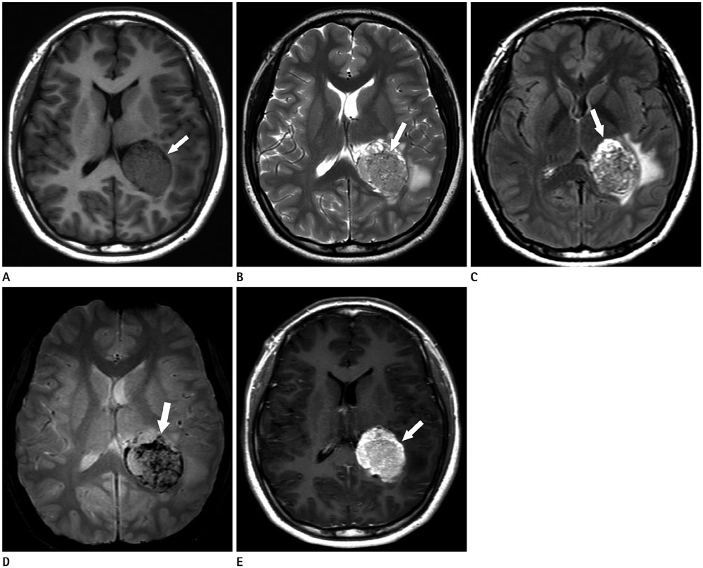
Fig. 2 MR images of intraventricular anaplastic hemangiopericytoma. A. On the axial T1-weighted image, the tumor (arrow) shows predominant isointensity with trigonal enlargement. B. This axial T2-weighted image reveals multiple, tiny vascular flow signal voids (arrow) within the tumor, with heterogeneous hyperintensity. C. Fluid-attenuated inversion recovery image shows a markedly hyperintense component (arrow) in the anterior portion of the tumor. D. On gradient echo image, countless hemorrhagic foci (arrow) can be seen in the tumor. E. Axial gadolinium contrast-enhanced T1-weighted image shows a lobular intraventricular tumor (arrow) with strong enhancement.

Fig. 3 Lateral left vertebral angiogram in a patient with intraventricular anaplastic hemangiopericytoma. A. In the arterial phase, the tumor margin is delineated by feeders, including the medial posterior choroidal artery (white arrows), the lateral posterior choroidal artery (short black arrows), and splenial branches of the posterior callosal artery (long black arrows). B. In the venous phase, the tumor shows more conspicuous staining (black double-lined arrows). There is a normal choroid plexus in front of the stained area of the tumor (white dashed arrows).
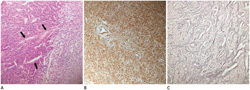
Fig. 4 Histopathologic examination of the anaplastic hemangiopericytoma (World Health Organization grade III). A. Photomicrographs shows dense cellularity and nuclear pleomorphism with slit-like vasculature (arrows) (original magnification, × 200; hematoxylin and eosin stain). B. Vimentin staining shows a cytoplasm-positive result (original magnification, × 200). C. Tumor cells stained positive for reticulin, highlighting blood vessels and pericellular reticulin (original magnification, × 200).
Reference
-
1. Muller J, Mealey J Jr. The use of tissue culture in differentiation between angioblastic meningioma and hemangiopericytoma. J Neurosurg. 1971; 34:341–348.2. Brunori A, Delitala A, Oddi G, Chiappetta F. Recent experience in the management of meningeal hemangiopericytomas. Tumori. 1997; 83:856–861.3. Rutkowski MJ, Sughrue ME, Kane AJ, Aranda D, Mills SA, Barani IJ, et al. Predictors of mortality following treatment of intracranial hemangiopericytoma. J Neurosurg. 2010; 113:333–339.4. Avinash KS, Thakar S, Ghosal N, Hegde AS. Anaplastic hemangiopericytoma in the frontal horn of the lateral ventricle. J Clin Neurosci. 2016; 26:147–149.5. Towner JE, Johnson MD, Li YM. Intraventricular hemangiopericytoma: a case report and literature review. World Neurosurg. 2016; 89:728.e5–728.e10.6. Desai K, Nadkarni T, Fattepurkar S, Goel A, Shenoy A, Chitale A, et al. Hemangiopericytoma in the trigone of the lateral ventricle--case report. Neurol Med Chir (Tokyo). 2004; 44:484–488.7. Chiechi MV, Smirniotopoulos JG, Mena H. Intracranial hemangiopericytomas: MR and CT features. AJNR Am J Neuroradiol. 1996; 17:1365–1371.8. Rutkowski MJ, Jian BJ, Bloch O, Chen C, Sughrue ME, Tihan T, et al. Intracranial hemangiopericytoma: clinical experience and treatment considerations in a modern series of 40 adult patients. Cancer. 2012; 118:1628–1636.9. Sumi K, Watanabe T, Ohta T, Fukushima T, Kano T, Yoshino A, et al. Hemangiopericytoma arising in the body of the lateral ventricle. Acta Neurochir (Wien). 2010; 152:145–149. discussion 150.10. Tanaka T, Kato N, Arai T, Hasegawa Y, Abe T. Hemangiopericytoma in the trigone of the lateral ventricle. Neurol Med Chir (Tokyo). 2011; 51:378–382.
- Full Text Links
-
- Actions
-
Cited
- CITED
-
- Close
- Share
-
- Similar articles
-
- Detection of Acute Intraventricular Hemorrhage: Comparison of FLAIR MR Imaging with Unenhanced CT
- CT and MRI Findings of Intraventricular Neurocytoma
- Comparison of Fluid-Attenuated Inversion-Recovery Magnetic Resonance Imaging with Computed Tomography in Acute Intraventricular Hemorrhage
- Magnetic resonance imaging of intracranial cysticercosis: comparison with computed tomography
- CT and Magnetic Resonance Imaging Findings of Lipomatous Hemangiopericytoma of Skull Base: A Case Report
