J Korean Ophthalmol Soc.
2016 Aug;57(8):1222-1227. 10.3341/jkos.2016.57.8.1222.
Changes in Choroidal Thickness in Branch Retinal Vein Occlusion
- Affiliations
-
- 1Department of Ophthalmology, Sanggye Paik Hospital, Inje University College of Medicine, Seoul, Korea. 991027js@hanmail.net
- 2Department of Ophthalmology, Seoul Paik Hospital, Inje University College of Medicine, Seoul, Korea.
- KMID: 2349067
- DOI: http://doi.org/10.3341/jkos.2016.57.8.1222
Abstract
- PURPOSE
To compare the choroidal thickness of a branch retinal vein occlusion (BRVO) lesion and that of other areas in the eyes.
METHODS
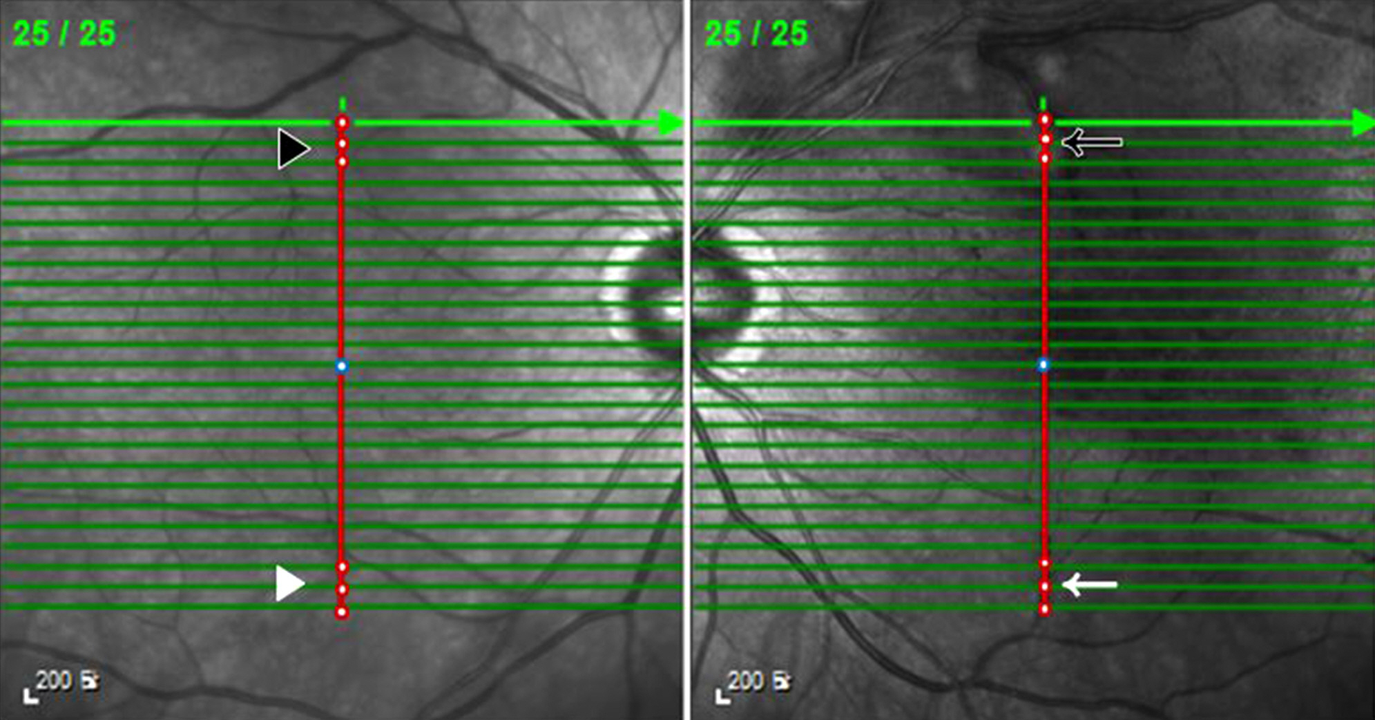
Patients who visited the Ophthalmologic Clinic of Inje University Sanggye Paik Hospital for BRVO between March 2015 and October 2015 were reviewed retrospectively. We performed basic ophthalmologic exam and enhanced depth imaging optical coherence tomography in 48 eyes of 24 patients with BRVO. The choroidal thickness was compared in a total of 4 places, the branch retinal vein occlusion lesion, the symmetric site in the same eye, and the equivalent sites in the fellow eye by paired t-test. All measurements were performed by 2 independent observers.
RESULTS
Choroidal thickness had strong inter-observer correlation. Choroidal thickness of the BRVO lesion was significantly thicker than that in the symmetric site of same eye, the equivalent site of lesion, and the equivalent site of the symmetric site to lesion in the fellow eye.
CONCLUSIONS
Choroidal thickness in acute BRVO lesions was thicker than choroidal thickness in other areas of the eyes. It is thought that both hydrostatic pressure and the effects of vascular endothelial growth factor influence choroidal thickness in the acute phase of BRVO.
Keyword
MeSH Terms
Figure
Reference
-
References
1. Weinberg D, Dodwell DG, Fern SA. Anatomy of arteriovenous crossings in branch retinal vein occlusion. Am J Ophthalmol. 1990; 109:298–302.
Article2. Christoffersen NL, Larsen M. Pathophysiology and hemodynamics of branch retinal vein occlusion. Ophthalmology. 1999; 106:2054–62.
Article3. Uemoto R, Nakasato-Sonn H, Kawagoe T, et al. Factors associated with enlargement of chorioretinal atrophy after intravitreal abdominal for myopic choroidal neovascularization. Graefes Arch Clin Exp Ophthalmol. 2012; 250:989–97.4. Tsuiki E, Suzuma K, Ueki R, et al. Enhanced depth imaging optical coherence tomography of the choroid in central retinal vein occlusion. Am J Ophthalmol. 2013; 156:543–7.e1.
Article5. Shin YU, Lee MJ, Lee BR. Choroidal maps in different types of macular edema in branch retinal vein occlusion using swept-source optical coherence tomography. Am J Ophthalmol. 2015; 160:328–34.e1.
Article6. Chung SE, Kang SW, Lee JH, Kim YT. Choroidal thickness in abdominal choroidal vasculopathy and exudative age-related macular degeneration. Ophthalmology. 2011; 118:840–5.7. Fujiwara T, Imamura Y, Margolis R, et al. Enhanced depth imaging optical coherence tomography of the choroid in highly myopic eyes. Am J Ophthalmol. 2009; 148:445–50.
Article8. Imamura Y, Fujiwara T, Margolis R, et al. Enhanced depth imaging optical coherence tomography of the choroid in central serous chorioretinopathy. Retina. 2009; 29:1469–73.
Article9. Maruko I, Iida T, Sugano Y, et al. Subfoveal choroidal thickness in fellow eyes of patients with central serous chorioretinopathy. Retina. 2011; 31:1603–8.
Article10. Maruko I, Iida T, Sugano Y, et al. Subfoveal choroidal thickness abdominal treatment of Vogt-Koyanagi-Harada disease. Retina. 2011; 31:510–7.11. Maruko I, Iida T, Sugano Y, et al. Subfoveal retinal and choroidal thickness after verteporfin photodynamic therapy for polypoidal choroidal vasculopathy. Am J Ophthalmol. 2011; 151:594–603.e1.
Article12. Du KF, Xu L, Shao L, et al. Subfoveal choroidal thickness in retinal vein occlusion. Ophthalmology. 2013; 120:2749–50.
Article13. Yuan A, Ahmad BU, Xu D, et al. Comparison of intravitreal abdominal and bevacizumab for the treatment of macular edema abdominal to retinal vein occlusion. Int J Ophthalmol. 2014; 7:86–91.14. Campochiaro PA, Sophie R, Pearlman J, et al. abdominal outcomes in patients with retinal vein occlusion treated with ranibizumab: the RETAIN study. Ophthalmology. 2014; 121:209–19.15. Aiello LP, Northrup JM, Keyt BA, et al. Hypoxic regulation of abdominal endothelial growth factor in retinal cells. Arch Ophthalmol. 1995; 113:1538–44.16. Ku DD, Zaleski JK, Liu S, et al. Vascular endothelial growth factor induces EDRF-dependent relaxation in coronary arteries. Am J Physiol. 1993; 265(2 Pt 2):H586–92.
Article17. Tilton RG, Chang KC, LeJeune WS, et al. Role for nitric oxide in the hyperpermeability and hemodynamic changes induced by abdominal VEGF. Invest Ophthalmol Vis Sci. 1999; 40:689–96.18. Ferrara N. Vascular endothelial growth factor: molecular and abdominal aspects. Curr Top Microbiol Immunol. 1999; 237:1–30.19. Jirarattanasopa P, Ooto S, Tsujikawa A, et al. Assessment of abdominal choroidal thickness by optical coherence tomography and abdominalgraphic changes in central serous chorioretinopathy. Ophthalmology. 2012; 119:1666–78.
- Full Text Links
- Actions
-
Cited
- CITED
-
- Close
- Share
- Similar articles
-
- OCT Biomarkers Predicting Recurrence of Macular Edema Secondary to Branch Retinal Vein Occlusion
- Clinical Features According to the Occlusion Site in Patients with Branch Retinal Vein Occlusion
- Clinical observation of the bilateral branch vein occlusion
- Retinal Arteriolar Changes in a Patient with Branch Retinal Vein Occlusion
- Eales Disease Accompanied with Branch Retinal Vein Occlusion


