Granular Cell Tumors on Unusual Anatomic Locations
- Affiliations
-
- 1Department of Dermatology, Severance Hospital, Yonsei University College of Medicine, Seoul, Korea. mglee@yuhs.ac
- KMID: 2345906
- DOI: http://doi.org/10.3349/ymj.2015.56.6.1731
Abstract
- Granular cell tumors (GCTs) are soft tissue tumors, which are thought to be derived from Schwann cells. Although most GCTs are reported to arise in tongue and oral cavity (30-50%), they can appear on any anatomic sites, even visceral organs. Herein, we report 5 cases of GCTs on unusual anatomic locations, such as palm, arm, thigh, finger, and vulvar area. Complete surgical excision is preferred treatment of choice to prevent recurrence. These cases emphasize that GCTs not involving oral cavity are more prevalent than expected, and the diagnosis should be histopathologically confirmed.
Keyword
MeSH Terms
Figure
-
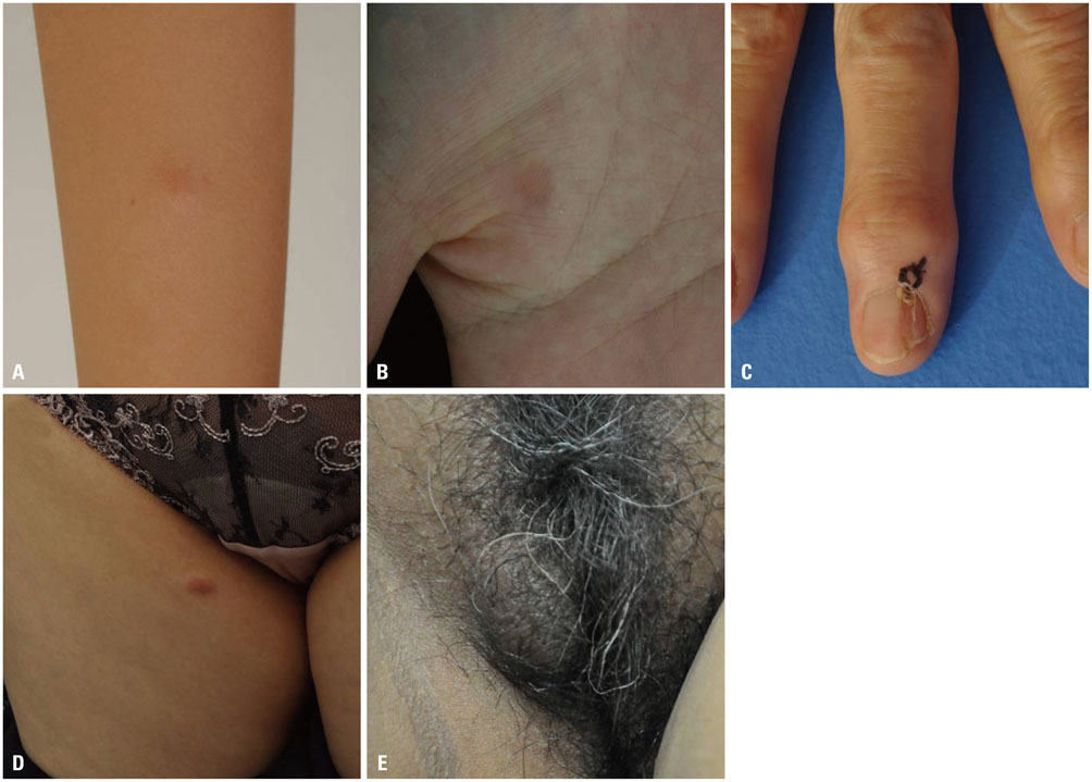
Fig. 1 (A) A solitary skin-colored pea-sized papule on right forearm of a 9-year-old female patient. (B) A bean-sized erythematous to brownish nodule on right palm of a 26-year-old female patient. (C) A longitudinal depression on nail plate with matchhead-sized dark pigmented crust on right third fingernail of a 69-year-old female patient. (D) A pea-sized light brown palpable firm nodule on the right thigh of a 52-year-old female patient. (E) Two non-tender nodules, each 3×1.5 cm and 1×1 cm sized, on the right labium majora of a 56-year-old female patient.

Fig. 2 Microphotographs of the tumors. (A) In case 1, a dermal tumor composed of fascicles, nests of large eosinophilic polygonal cells with granular cytoplasm (H&E, ×400). (B) Tumor cells were positive for S-100 immunostain (S-100, ×400). (C) The tumor cells of case 2 contain abundant eosinophilic cytoplasm with fine granules, suggesting granular cell tumor (H&E, ×400). (D) Immunhistochemical stain with S-100 was positive in tumor cells (S-100, ×400). (E) Nail matrix biopsy of the case 3 revealed proliferations of round cells with abundant eosinophilic cytoplasm (H&E, ×400). (F) Immunhistochemical stain with S-100 was positive in tumor cells (S-100, ×400). (G) Histopathologic evaluation of the tumor in case 4 shows tumor cells with eosinophilic granular cytoplasm (H&E, ×400). (H) S-100 was positively stained in tumor cells (S-100, ×400). Microphotographs of the tumors. (I) Infiltrating sheets of tumor cells with eosinophilic granular cytoplasm, consistent with granular cell tumor (H&E, ×400). (J) The cells were positive for S-100 staining (S-100, ×400).
Reference
-
1. Lack EE, Worsham GF, Callihan MD, Crawford BE, Klappenbach S, Rowden G, et al. Granular cell tumor: a clinicopathologic study of 110 patients. J Surg Oncol. 1980; 13:301–316.
Article2. Newman E, Eichenfield LF. Granular cell tumor on the palm of an 8-year-old girl. Pediatr Dermatol. 2010; 27:656–657.
Article3. de Misa RF, Castro V, Suárez J, Perera A. Pruritic vulvar nodule in a black woman. Diagnosis: Granular cell tumor (Abrikossoff tumor). Arch Dermatol. 2000; 136:1165–1170.4. Hong SC, Lim YK, Chew SH, Chia YN, Yam KL. Case report of granular cell tumor of the vulva and review of current literature. Gynecol Oncol Case Rep. 2012; 3:20–22.
Article5. Rosai J, Ackerman LV. Rosai and Ackerman's surgical pathology. 9th ed. Edinburgh: Mosby;2004.6. Apisarnthanarax P. Granular cell tumor. An analysis of 16 cases and review of the literature. J Am Acad Dermatol. 1981; 5:171–182.7. Ramos PC, Kapp DS, Longacre TA, Teng NN. Malignant granular cell tumor of the vulva in a 17-year-old: case report and literature review. Int J Gynecol Cancer. 2000; 10:429–434.
Article8. Torrijos-Aguilar A, Alegre-de Miquel V, Pitarch-Bort G, Mercader-García P, Fortea-Baixauli JM. [Cutaneous granular cell tumor: a clinical and pathologic analysis of 34 cases]. Actas Dermosifiliogr. 2009; 100:126–132.
Article9. Fanburg-Smith JC, Meis-Kindblom JM, Fante R, Kindblom LG. Malignant granular cell tumor of soft tissue: diagnostic criteria and clinicopathologic correlation. Am J Surg Pathol. 1998; 22:779–794.10. Cheewakriangkrai C, Sharma S, Deeb G, Lele S. A rare female genital tract tumor: benign granular cell tumor of vulva: case report and review of the literature. Gynecol Oncol. 2005; 97:656–658.
Article11. Levavi H, Sabah G, Kaplan B, Tytiun Y, Braslavsky D, Gutman H. Granular cell tumor of the vulva: six new cases. Arch Gynecol Obstet. 2006; 273:246–249.
Article12. Horowitz IR, Copas P, Majmudar B. Granular cell tumors of the vulva. Am J Obstet Gynecol. 1995; 173:1710–1713.
Article
