Ann Dermatol.
2016 Aug;28(4):517-519. 10.5021/ad.2016.28.4.517.
Acral Persistent Papular Mucinosis with Partial Response to Tacrolimus Ointment
- Affiliations
-
- 1Department of Dermatology, Samsung Medical Center, Sungkyunkwan University School of Medicine, Seoul, Korea. dylee@skku.edu
- KMID: 2344833
- DOI: http://doi.org/10.5021/ad.2016.28.4.517
Abstract
- No abstract available.
MeSH Terms
Figure
-
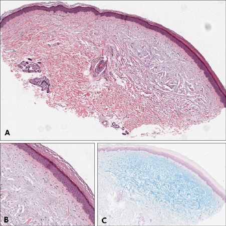
Fig. 1 (A) Epidermis is normal. Deposition of bluish material is seen at upper to mid dermis, fairly discrete (H&E, ×20). (B) Small grenz zone was spared. Fibroblasts were present among the deposition but not seem to be proliferated. Inflammatory cell infiltration in dermis is barely seen (H&E, ×100). (C) The bluish material was positive with alcian blue (Alcian blue pH 2.5, ×20).
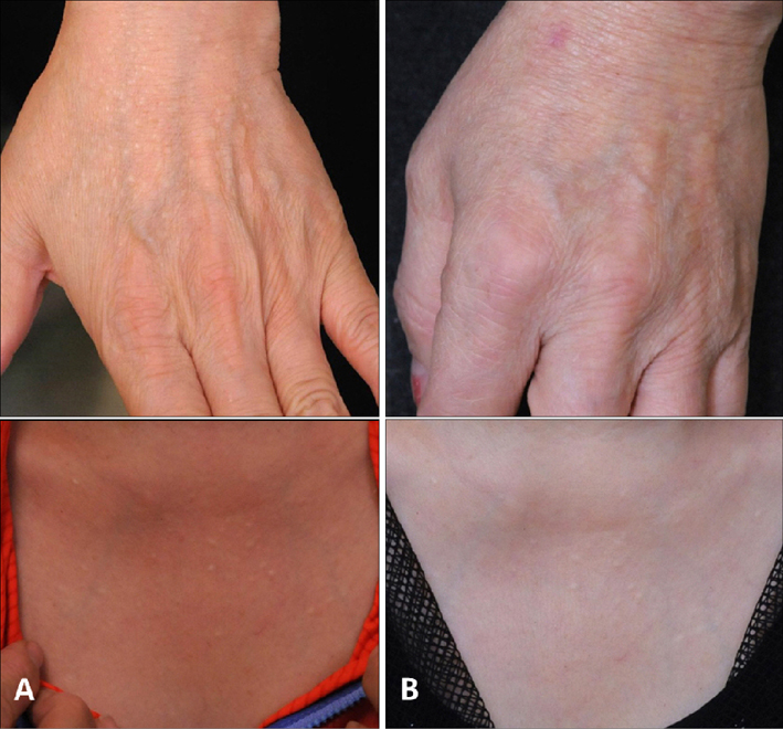
Fig. 2 (A) Left dorsum of hand and anterior chest before treatment. (B) Left dorsum of hand and anterior chest after 15 weeks of tacrolimus ointment 0.1% application.
Reference
-
1. Luo DQ, Wu LC, Liu JH, Zhang HY. Acral persistent papular mucinosis: a case report and literature review. J Dtsch Dermatol Ges. 2011; 9:354–359.
Article2. André Jorge F, Mimura Cortez T, Guadalini Mendes F, Esther Alencar Marques M, Amante Miot H. Treatment of acral persistent papular mucinosis with electrocoagulation. J Cutan Med Surg. 2011; 15:227–229.
Article3. Song JY, Lee SW, Kim CW, Kim HO. A case of acral persistent papular mucinosis. Ann Dermatol. 2002; 14:178–180.
Article4. Harris JE, Purcell SM, Griffin TD. Acral persistent papular mucinosis. J Am Acad Dermatol. 2004; 51:982–988.
Article5. Rongioletti F, Zaccaria E, Cozzani E, Parodi A. Treatment of localized lichen myxedematosus of discrete type with tacrolimus ointment. J Am Acad Dermatol. 2008; 58:530–532.
Article
