J Korean Soc Radiol.
2011 Nov;65(5):479-485.
The Image Quality of a Digital Chest X-Ray Radiography System: Comparison of Quantitative Image Quality Analysis and Radiologists' Visual Scoring
- Affiliations
-
- 1Department of Radiation Oncology, Pusan National University Yangsan Hospital, Yangsan, Korea.
- 2Department of Radiology, Samsung Medical Center, Seoul, Korea. chungmjmd@gmail.com
- 3Department of Radiation Oncology, Pusan National University Hospital, Busan, Korea.
- 4Research Institute for Convergence of Biomedical Science and Technology, Pusan National University Yangsan Hospital, Yangsan, Korea.
Abstract
- PURPOSE
To evaluate the performance of imaging devices, which should be periodically monitored to maintain high quality images to the radiologists. Additionally, this evaluation may prevent patients from radiation over-exposure. The most suitable engineering standard for imaging performance evaluation of digital X-ray thoracic imagers was determined.
MATERIALS AND METHODS
IEC 62220-1 standards were used to evaluate the performance of the imagers. In succession, the visibilities of overall image, pneumothorax, and humerus head in anthropomorphic thoracic phantom images were used to evaluate the image qualities by radiologists.
RESULTS
The rank correlation coefficient (p) of visual scoring by radiologists with system spatial resolution is not meaningful (p-value, p = 0.295), but is significant with image noise (p-value, p = -0.9267). Finally, the noise equivalent quanta (NEQ) presents a high rank correlation for visual scoring of radiologists (p-value, p = 0.9320).
CONCLUSION
Image quality evaluation of radiologists were mainly affected by imaging noise. Hence, the engineered standard for evaluating image noise is the most important index to effectively monitor the performance of X-ray imagers. Additionally, the NEQ can be used to evaluate the performance of radiographic systems, because it theoretically corresponds to the synthetic image quality of systems.
MeSH Terms
Figure
-

Fig. 1 An example of acquired images for MTF and NNPS measurements. Note.-MTF = modulation transfer function, NNPS = normalized noise power spectrum
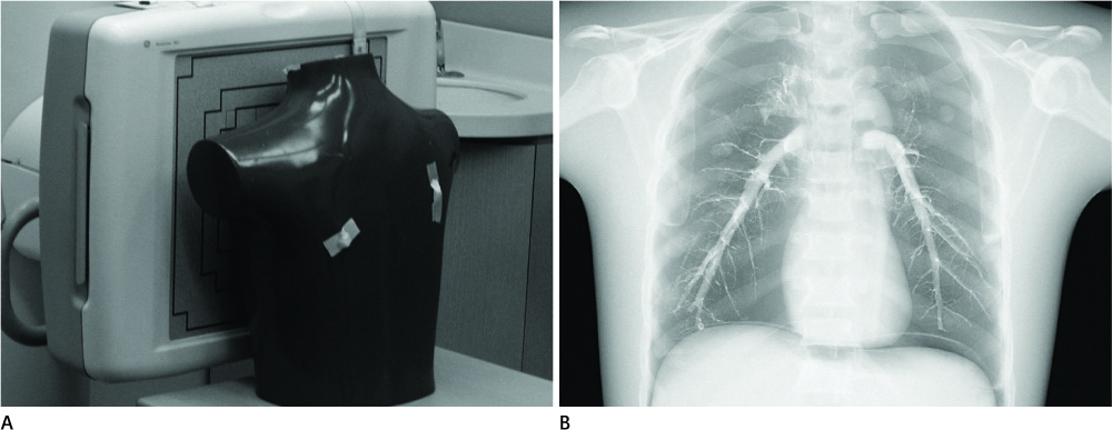
Fig. 2 (A) An anthropomorphic phantom and (B) its radiographic image.
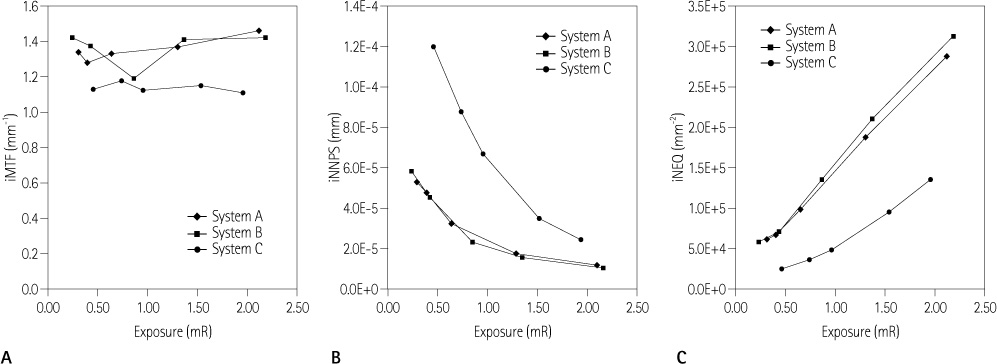
Fig. 3 Relationships between exposures and (A) iMTFs, (B) iNNPSs and (C) iNEQs. Note.-iMTF = integrated modulation transfer function, iNEQ = integrated noise equivalent quanta, iNNPS = integrated normalized noise power spectrum

Fig. 4 Relationships between mean scores by radiologists and (A) exposures, (B) iMTFs, (C) iNNPSs, and (D) iNEQs. Note.-iMTF = integrated modulation transfer function, iNEQ = integrated noise equivalent quanta, iNNPS = integrated normalized noise power spectrum
Reference
-
1. Stierstorfer K, Spahn M. Self-normalizing method to measure the detective quantum efficiency of a wide range of x-ray detectors. Med Phys. 1999; 26:1312–1319.2. Hillen W, Schiebel U, Zaengel T. Imaging performance of a digital storage phosphor system. Med Phys. 1987; 14:744–751.3. Cunningham IA. Standard for measurement of noise power spectra. AAPM Report. 1999.4. Samei E, Flynn MJ, Reimann DA. A method for measuring the presampled MTF of digital radiographic systems using an edge test device. Med Phys. 1998; 25:102–113.5. ICRU. Medical imaging - the assessment of image quality. ICRU Report. 1996. 54.6. IEC. Medical electrical equipment - characteristics of digital X-ray imaging devices - determination of the detective quantum efficiency. IEC 62220-1. 2001.7. Herrmann C, Sund P, Tingberg A, Almen A, Mattsson S, Keddache S, et al. Comparison of two methods for evaluating image quality of chest radiographs. Proc SPIE. 2000; 251:3981–3981.8. Sund P, Båth M, Kheddache S, Månsson LG. Comparison of visual grading analysis and determination of detective quantum efficiency for evaluating system performance in digital chest radiography. Eur Radiol. 2004; 14:48–58.9. Cunningham IA. Metrics of system performance. SPIE PRESS;2000. p. 122–123.10. European Commission. European guidelines on quality criteria for diagnostic radiographic images. EUR. 1996. p. 16260.11. Borasi G, Nitrosi A, Ferrari P, Tassoni D. On site evaluation of three flat panel detectors for digital radiography. Med Phys. 2003; 30:1719–1731.12. Samei E, Flynn MJ. An experimental comparison of detector performance for direct and indirect digital radiography systems. Med Phys. 2003; 30:608–622.13. Ehsan S, Michael JF, Chotas HG, Dobbins JT. DQE of direct and indirect digital radiographic systems. Proc SPIE. 2001; 4:189–197.
- Full Text Links
-
- Actions
-
Cited
- CITED
-
- Close
- Share
-
- Similar articles
-
- Objective and Quantitative Evaluation of Image Quality Using Fuzzy Integral: Phantom Study
- A Comparative Study of Image Quality and Radiation Dose with Changes in Tube Voltage and Current for a Digital Chest Radiography
- Storage Phosphor Digital Radiography in Portable Chest Imaging: Comparison of Image Quality with Conventional Film-Screen System with Variation of mAs
- Relationship of Image Quality and Radiation Dose for Chest Radiography in the Medical Institutions for Pneumoconiosis: A Comparison to the Korean Diagnostic Reference Level
- Image Enhancement and Clinical Evaluation in Digital Chest Radiography
