Granular Cell Tumor of the Neurohypophysis: A Case Report with Magnetic Resonance and CT Imaging Findings
- Affiliations
-
- 1Department of Radiology, Seoul St. Mary's Hospital, The Catholic University of Korea College of Medicine, Seoul, Korea. ahn-kj@catholic.ac.kr
- 2Department of Pathology, Seoul St. Mary's Hospital, The Catholic University of Korea College of Medicine, Seoul, Korea.
- 3Department of Neurosurgery, Seoul St. Mary's Hospital, The Catholic University of Korea College of Medicine, Seoul, Korea.
Abstract
- A granular cell tumor (GCT) usually occurs as a small, solitary, nodular tumor and is more prevalent in adult females. The authors report the magnetic resonance (MR) and CT imaging findings in a 61-year-old woman with GCT of the neurohypophysis presenting with a history of reduced visual acuity in her right eye. MR images showed a suprasellar mass with an isointense signal on a T1-weighted image and an hypointense signal on a T2-weighted image. The histopathological examination revealed a granular cell tumor. In this article, the MR and CT imaging findings of GCT of the neurohypophysis with the literature reviews are discussed.
MeSH Terms
Figure
-
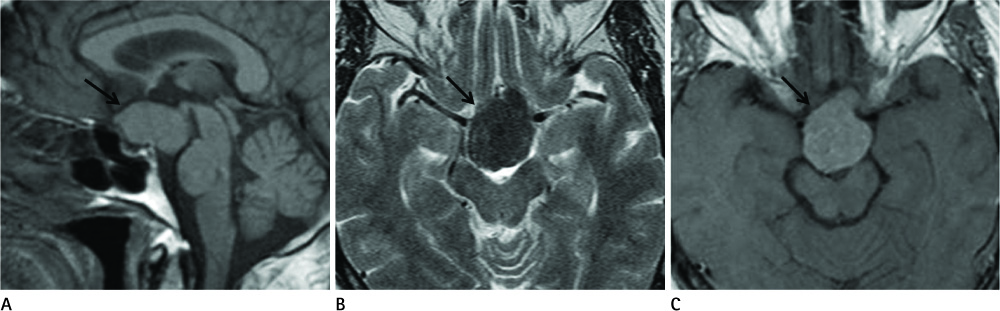
Fig. 1 A 61-year-old woman with a history of reduced visual acuity in her right eye for a period of 3 months. A. Sagittal T1-weighted image showing a large, well defined, smooth marginated mass (arrow) in the pituitary gland showing an isointense signal with the loss of normal posterior lobe hyperintensity. B. Axial T2-weighted image indicatinging that this mass (arrow) shows a hypointense signal. C. Contrast-enhanced axial T1-weighted image showing mild contrast enhancement is noted in the mass (arrow).
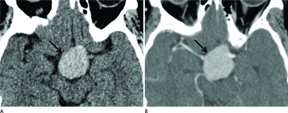
Fig. 2 CT on admission. A. On the non-enhanced CT image, this mass (arrow) is homogenous and shows a slightly higher density (about 53 HU). B. On the contrast-enhanced CT image, this mass (arrow) shows homogenous contrast enhancement.
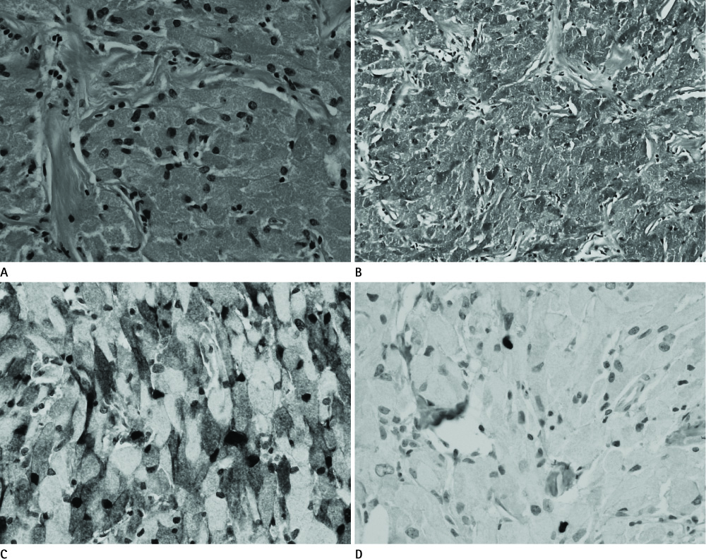
Fig. 3 Histological examination. A. Polygonal tumor cells are shown with abundant eosinophilic granular cytoplasm (H&E × 40). B. The cytoplasmic granules show positive staining in periodic acid-Schiff staining (× 100). C. Immunohistochemical staining for S-100 protein shows positive results (× 400). D. Ki-67 labeling index is less than 1% (× 400).
Reference
-
1. Schaller B, Kirsch E, Tolnay M, Mindermann T. Symptomatic granular cell tumor of the pituitary gland: case report and review of the literature. Neurosurgery. 1998; 42:166–170. discussion 170-171.2. Iglesias A, Arias M, Brasa J, Paramo C, Conde C, Fernandez R. MR imaging findings in granular cell tumor of the neurohypophysis: a difficult preoperative diagnosis. Eur Radiol. 2000; 10:1871–1873.3. Cone L, Srinivasan M, Romanul FC. Granular cell tumor (choristoma) of the neurohypophysis: two cases and a review of the literature. AJNR Am J Neuroradiol. 1990; 11:403–406.4. Barrande G, Kujas M, Gancel A, Turpin G, Bruckert E, Kuhn JM, et al. [Granular cell tumors. Rare tumors of the neurohypophysis]. Presse Med. 1995; 24:1376–1380.5. Landolt AM. Ultrastructure of human sella tumors. Correlations of clinical findings and morphology. Acta Neurochir (Wien). 1975; Suppl 22. 1–167.6. Bubl R, Hugo HH, Hempelmann RG, Barth H, Mehdorn HM. Granular-cell tumour: a rare suprasellar mass. Neuroradiology. 2001; 43:309–312.7. Ji CH, Teng MM, Chang T. Granular cell tumour of the neurohypophysis. Neuroradiology. 1995; 37:451–452.8. Boecher-Schwarz HG, Fries G, Bornemann A, Ludwig B, Perneczky A. Suprasellar granular cell tumor. Neurosurgery. 1992; 31:751–754. discussion 754.9. Becker DH, Wilson CB. Symptomatic parasellar granular cell tumors. Neurosurgery. 1981; 8:173–180.
- Full Text Links
-
- Actions
-
Cited
- CITED
-
- Close
- Share
-
- Similar articles
-
- Granular Cell Tumor of the Presacral Space: A Case Report
- CT and MR Imaging Findings of a Granular Cell Tumor of the Orbit: Report of Two Cases
- Granular Cell Tumor of the Urinary Bladder: A Case Report
- Granular Cell Tumor Simulating Breast Malignancy: A Case Report
- Granular Cell Tumor in the Pituitary Stalk: A Case Report
