Korean Circ J.
2016 Jul;46(4):580-583. 10.4070/kcj.2016.46.4.580.
Conservative Management of Left Atrial Intramural Hematoma after Catheter Ablation
- Affiliations
-
- 1Department of Echocardiography, Day General Hospital, Tehran, Iran. faraviolet@yahoo.com
- 2Department of Electrophysiology, Day General Hospital, Tehran, Iran.
- 3Department of Radiology, Day General Hospital, Tehran, Iran.
- KMID: 2344436
- DOI: http://doi.org/10.4070/kcj.2016.46.4.580
Abstract
- Left atrial intramural hematoma is a very rare complication of radiofrequency ablation procedures. A patient with tachyarrhythmia underwent radiofrequency catheter ablation. Echocardiography performed the following morning showed a large mass in the left atrium, suggestive of intramural hematoma formation. The patient was in a stable condition; therefore, it was decided that follow-up should be conservative and her anticoagulation therapy was continued. The size of the hematoma decreased significantly over the following 50 days. This case highlights a rare complication of a complex catheter ablation procedure in the left atrium that was managed via a noninvasive approach, with which all interventionists should be familiar.
Keyword
MeSH Terms
Figure
-
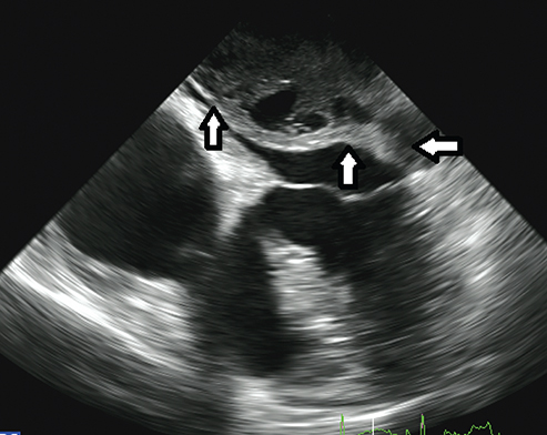
Fig. 1 A 4-chamber view of the transesophagial echocardiograph showing a large multi-loculated mass occupying most of the left atrial space (arrows) and partially obstructing the left ventricular inflow

Fig. 2 A computed tomographic angiography image showing a homogeneous non-enhancing intramural mass located in the posterosuperior left atrial wall (arrows).
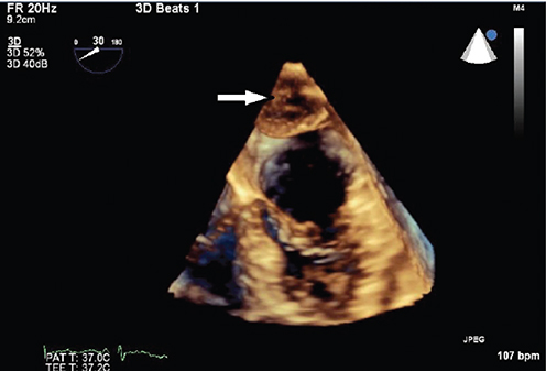
Fig. 3 A follow-up three-dimensional transesophagial echocardiograph (4-chamber view; in diastole) taken 50 days after discharge showing significant resolution of the left atrial hematoma (arrow).
Reference
-
1. Lanfranchi A, Gelpi G, Rossi RS, Lemma M. A fast-growing obstructive left atrial intramural hematoma causing acute prolonged chest pain. Interact Cardiovasc Thorac Surg. 2009; 9:363–365.2. Fukuhara S, Dimitrova KR, Geller CM, Hoffman DM, Tranbaugh RF. Left atrial dissection: an almost unknown entity. Interact Cardiovasc Thorac Surg. 2015; 20:96–100.3. Shaikh N, Rehman NU, Salazar MF, Grodman RS. Spontaneous intramural atrial hematoma presenting as a left atrial mass. J Am Soc Echocardiogr. 1999; 12:1101–1103.4. Gual-Capllonch F, Arce J, Serés L, et al. Left atrial intramural haematoma associated with mitral annular calcification. Eur J Echocardiogr. 2010; 11:E18.5. Kurek C, Gwechenberger M, Richter B, Binder T, Loewe C, Gössinger H. Intramural left atrial haematoma mimicking cardiac tamponade after catheter ablation of atrial fibrillation. Europace. 2009; 11:667–668.6. Kelly S, Bicknell SG, Sharma S. Left atrial wall hematoma after radiofrequency ablation for atrial fibrillation. AJR Am J Roentgenol. 2006; 186:1317–1319.7. Sah R, Epstein LM, Kwong RY. Images in cardiovascular medicine. Intramural atrial hematoma after catheter ablation for atrial tachyarrhythmias. Circulation. 2007; 115:e446–e447.
- Full Text Links
-
- Actions
-
Cited
- CITED
-
- Close
- Share
-
- Similar articles
-
- Left atrial ıntramural hematoma after radiofrequency catheter ablation of left lateral accessory pathway
- Management of Atrial Flutter
- Underdevelopment of Left Atrial Appendage
- Large Circular Ring Catheter Ablation Versus Anatomically Guided Ablation of Atrial Fibrillation: Back to the Future for Successful Catheter Ablation of Atrial Fibrillation?
- A Case of Successful Ablation of Right-Sided Accessory Pathway during Atrial Fibrillation
