Efficacy of Intravitreal Anti-vascular Endothelial Growth Factor or Steroid Injection in Diabetic Macular Edema According to Fluid Turbidity in Optical Coherence Tomography
- Affiliations
-
- 1HanGil Eye Hospital, Incheon, Korea. Jhsohn19@hanafos.com
Abstract
- PURPOSE
To determine if short term effects of intravitreal anti-vascular endothelial growth factor or steroid injection are correlated with fluid turbidity, as detected by spectral domain optical coherence tomography (SD-OCT) in diabetic macular edema (DME) patients.
METHODS
A total of 583 medical records were reviewed and 104 cases were enrolled. Sixty eyes received a single intravitreal bevacizumab injection (IVB) on the first attack of DME and 44 eyes received triamcinolone acetonide treatment (IVTA). Intraretinal fluid turbidity in DME patients was estimated with initialintravitreal SD-OCT and analyzed with color histograms from a Photoshop program. Central macular thickness and visual acuity using a logarithm from the minimum angle of resolution chart, were assessed at the initial period and 2 months after injections.
RESULTS
Visual acuity and central macular thickness improved after injections in both groups. In the IVB group, visual acuity and central macular thickness changed less as the intraretinal fluid became more turbid. In the IVTA group, visual acuity underwent less change while central macular thickness had a greater reduction (r = -0.675, p = 0.001) as the intraretinal fluid was more turbid.
CONCLUSIONS
IVB and IVTA injections were effective in reducing central macular thickness and improving visual acuity in DME patients. Further, fluid turbidity, which was detected by SD-OCT may be one of the indexes that highlight the influence of the steroid-dependent pathogenetic mechanism.
Keyword
MeSH Terms
-
Aged
Angiogenesis Inhibitors/*therapeutic use
Bevacizumab/*therapeutic use
Diabetic Retinopathy/*drug therapy/physiopathology
Female
Glucocorticoids/*therapeutic use
Humans
Intravitreal Injections
Macular Edema/*drug therapy/physiopathology
Male
Middle Aged
Nephelometry and Turbidimetry
Retina/pathology
*Subretinal Fluid
Tomography, Optical Coherence
Treatment Outcome
Triamcinolone Acetonide/*therapeutic use
Vascular Endothelial Growth Factor A/antagonists & inhibitors
Visual Acuity/physiology
Angiogenesis Inhibitors
Bevacizumab
Glucocorticoids
Triamcinolone Acetonide
Vascular Endothelial Growth Factor A
Figure
-
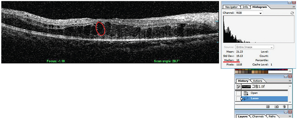
Fig. 1 The measurement of intraretinal fluid turbidity. Fluid turbidity was measured using a Photoshop program. Every cystoid area in intraretinal layer was dragged using the Photoshop dragging tool and a tablet pen. The color histogram of every dragged image was examined and the median value of each image was obtained.
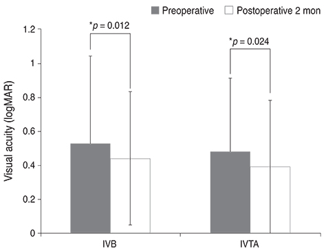
Fig. 2 Change of the mean best-corrected visual acuity after intravitreal injection. The best-corrected visual acuity was expressed as a logarithm of the minimum angle of the resolution chart (logMAR). After a period of 2 months after intravitreal injection, both intravitreal bevacizumab injection (IVB) and intravitreal triamcinolone acetonide injection (IVTA) groups showed statistically significant improvement in visual acuity (p = 0.0012 in IVB and p = 0.0024 in IVTA, respectively). *Statistically significant (p < 0.05) by Mann-Whitney test.
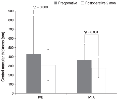
Fig. 3 Change of central macular thickness after intravitreal injection. At the 2 month postoperative time point central macular thickness was significantly reduced after intravitreal injection in intravitreal bevacizumab injection (IVB) and intravitreal triamcinolone acetonide injection (IVTA) groups (p = 0.000 in IVB and p = 0.0001 in IVTA, respectively). *Statistically significant (p < 0.05) by Mann-Whitney test.
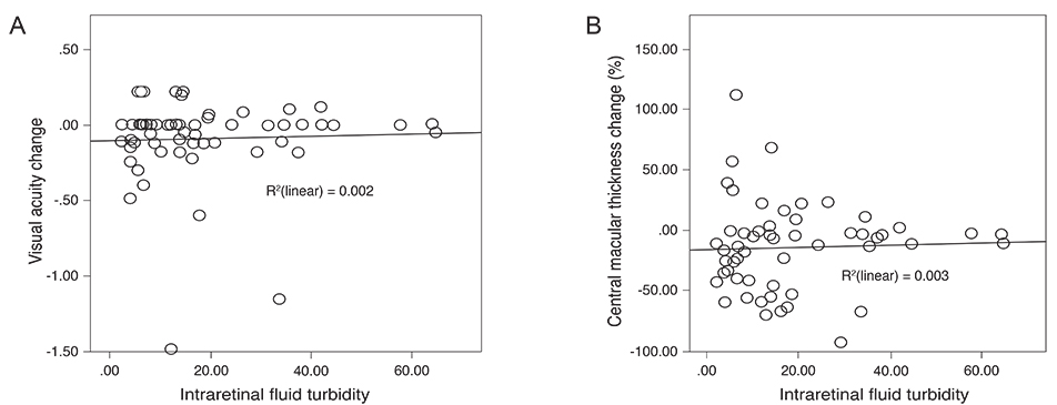
Fig. 4 Correlation between intraretinal fluid turbidity and the change of both the mean best-corrected visual acuity (A) and central macular thickness (CMT) (B) in the intravitreal bevacizumab injection (IVB) group. Change of the best mean corrected visual acuity is defined as the subtraction between initial best-corrected visual acuity and the 2 month postoperative best-corrected visual acuity. Further the change of CMT is defined as [(CMTat baseline - CMTat postoperative 2 months) / CMTat baseline] × 100. As the intraretinal fluid turbidity increased in the IVB group, the best-corrected visual acuity (A) and CMT (B) remained unchanged compared to initial measurements.
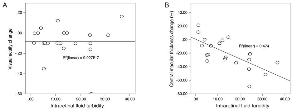
Fig. 5 Change of the mean best-corrected visual acuity (A) and central macular thickness (CMT) (B) according to the intraretinal fluid turbidity in the intravitreal triamcinolone acetonide injection (IVTA) group. Change of mean best-corrected visual acuity is defined as the subtraction between initial best-corrected visual acuity and the 2 month postoperative best-corrected visual acuity. The change of CMT is defined as [(CMTat baseline - CMTat postoperative 2 months) / CMTat baseline] × 100. In the IVTA group, as the intraretinal fluid turbidity increased, the best-corrected visual acuity was unchanged in comparison with those of initial measurements (A), and the CMT reduced greatly (B).
Reference
-
1. Klein R, Klein BE, Moss SE. Visual impairment in diabetes. Ophthalmology. 1984; 91:1–9.2. Moss SE, Klein R, Klein BE. Ten-year incidence of visual loss in a diabetic population. Ophthalmology. 1994; 101:1061–1070.3. Antcliff RJ, Marshall J. The pathogenesis of edema in diabetic maculopathy. Semin Ophthalmol. 1999; 14:223–232.4. Montero JA, Ruiz-Moreno JM, De La Vega C. Incomplete posterior hyaloid detachment after intravitreal pegaptanib injection in diabetic macular edema. Eur J Ophthalmol. 2008; 18:469–472.5. Ahmadieh H, Ramezani A, Shoeibi N, et al. Intravitreal bevacizumab with or without triamcinolone for refractory diabetic macular edema; a placebo-controlled, randomized clinical trial. Graefes Arch Clin Exp Ophthalmol. 2008; 246:483–489.6. Goyal S, Lavalley M, Subramanian ML. Meta-analysis and review on the effect of bevacizumab in diabetic macular edema. Graefes Arch Clin Exp Ophthalmol. 2011; 249:15–27.7. Soheilian M, Ramezani A, Bijanzadeh B, et al. Intravitreal bevacizumab (avastin) injection alone or combined with triamcinolone versus macular photocoagulation as primary treatment of diabetic macular edema. Retina. 2007; 27:1187–1195.8. Soheilian M, Ramezani A, Obudi A, et al. Randomized trial of intravitreal bevacizumab alone or combined with triamcinolone versus macular photocoagulation in diabetic macular edema. Ophthalmology. 2009; 116:1142–1150.9. Lim JW, Lee HK, Shin MC. Comparison of intravitreal bevacizumab alone or combined with triamcinolone versus triamcinolone in diabetic macular edema: a randomized clinical trial. Ophthalmologica. 2012; 227:100–106.10. Song JH, Lee JJ, Lee SJ. Comparison of the short-term effects of intravitreal triamcinolone acetonide and bevacizumab injection for diabetic macular edema. Korean J Ophthalmol. 2011; 25:156–160.11. Paccola L, Costa RA, Folgosa MS, et al. Intravitreal triamcinolone versus bevacizumab for treatment of refractory diabetic macular oedema (IBEME study). Br J Ophthalmol. 2008; 92:76–80.12. Shimura M, Yasuda K, Nakazawa T, et al. Visual outcome after intravitreal triamcinolone acetonide depends on optical coherence tomographic patterns in patients with diffuse diabetic macular edema. Retina. 2011; 31:748–754.13. Kim M, Lee P, Kim Y, et al. Effect of intravitreal bevacizumab based on optical coherence tomography patterns of diabetic macular edema. Ophthalmologica. 2011; 226:138–144.14. Wu PC, Lai CH, Chen CL, Kuo CN. Optical coherence tomographic patterns in diabetic macula edema can predict the effects of intravitreal bevacizumab injection as primary treatment. J Ocul Pharmacol Ther. 2012; 28:59–64.15. Nguyen QD, Shah SM, Heier JS, et al. Primary end point (six months) results of the ranibizumab for edema of the mAcula in diabetes (READ-2) study. Ophthalmology. 2009; 116:2175–2181.16. Nguyen QD, Shah SM, Khwaja AA, et al. Two-year outcomes of the ranibizumab for edema of the mAcula in diabetes (READ-2) study. Ophthalmology. 2010; 117:2146–2151.17. Mitchell P, Bandello F, Schmidt-Erfurth U, et al. The RESTORE study: ranibizumab monotherapy or combined with laser versus laser monotherapy for diabetic macular edema. Ophthalmology. 2011; 118:615–625.18. Nguyen QD, Brown DM, Marcus DM, et al. Ranibizumab for diabetic macular edema: results from 2 phase III randomized trials: RISE and RIDE. Ophthalmology. 2012; 119:789–801.19. Michaelides M, Kaines A, Hamilton RD, et al. A prospective randomized trial of intravitreal bevacizumab or laser therapy in the management of diabetic macular edema (BOLT study) 12-month data: report 2. Ophthalmology. 2010; 117:1078–1086.20. Diabetic Retinopathy Clinical Research Network. Scott IU, Edwards AR, et al. A phase II randomized clinical trial of intravitreal bevacizumab for diabetic macular edema. Ophthalmology. 2007; 114:1860–1867.21. Funatsu H, Yamashita H, Noma H, et al. Increased levels of vascular endothelial growth factor and interleukin-6 in the aqueous humor of diabetics with macular edema. Am J Ophthalmol. 2002; 133:70–77.22. Sohn HJ, Han DH, Kim IT, et al. Changes in aqueous concentrations of various cytokines after intravitreal triamcinolone versus bevacizumab for diabetic macular edema. Am J Ophthalmol. 2011; 152:686–694.23. Browning DJ, Apte RS, Bressler SB, et al. Association of the extent of diabetic macular edema as assessed by optical coherence tomography with visual acuity and retinal outcome variables. Retina. 2009; 29:300–305.
- Full Text Links
-
- Actions
-
Cited
- CITED
-
- Close
- Share
-
- Similar articles
-
- Current Concepts in Diabetic Retinopathy
- Intravitreal and Additional Posterior Subtenon Triamcinolone Injection in Diabetic Macular Edema
- The Effect of Intravitreal Triamcinolone Acetonide Injection according to the Diabetic Macular Edema Type
- A Multimodal Approach to Diabetic Macular Edema
- Ganglion Cell Layer Thickness after Anti-Vascular Endothelial Growth Factor Treatment in Retinal Vein Occlusion
