A Case of Maculopathy from Handheld Green Laser Pointer
- Affiliations
-
- 1Department of Ophthalmology, Gyeongsang National University School of Medicine, Jinju, Korea. medcabin@hanmail.net
- 2Gyeongsang Institute of Health Science, Gyeongsang National University, Jinju, Korea.
- KMID: 2339054
- DOI: http://doi.org/10.3341/jkos.2015.56.3.447
Abstract
- PURPOSE
To report a case of successfully treated maculopathy after exposure to a handheld green laser pointer beam.
CASE SUMMARY
A-15-year-old patient visited our clinic complaining of visual disturbance in the left eye 5 days earlier after exposure to a handheld laser pointer with 532 nm wavelength green beam for 5 seconds. His best corrected visual acuity was 20/50 in the left eye. On fundus examination, a yellowish retinal scar was observed at the foveal area. The spectral domain optical coherence tomography (SD-OCT) showed cone outer segment tip line and inner segment/outer segment line disruption, external limiting membrane line and retinal pigment epithelial complex injury related to laser pointer exposure. We started occlusion therapy, oral prednisolone and, antioxidant treatment on his left eye for 2 weeks. The best corrected visual acuity was 20/20 in the left eye at 1 month after treatment. However, spectral domain optical coherence tomography showed a scar remained in the retinal pigment epithelial complex of the macular region of his left eye while the external limiting membrane line was restored and inner segment/outer segment line was partially restored.
CONCLUSIONS
Maculopathy can result from exposure to a handheld green laser pointer. Occlusion therapy, oral prednisolone and, antioxidant treatment might be helpful for recovery of visual acuity and restoration of external limiting membrane line.
Keyword
MeSH Terms
Figure
-

Figure 1. Fundus photograph (A) and SD-OCT (B) at the first ophthalmic examination show yellowish retinal scar at the foveal area and cone outer segment tip line and inner segment/outer segment line disruption, external limiting membrane line and retinal pigment epithelial complex injury in the left eye. SD-OCT = spectral domain optical coherence tomography.

Figure 2. Fundus photograph (A) and SD-OCT (B) after 2 weeks of treatment show external limiting membrane, inner segment/outer segment line were partially restored, but retinal scar at the foveal area and retinal pigment epithelial complex injury persist in the left eye. SD-OCT = spectral domain optical coherence tomography.
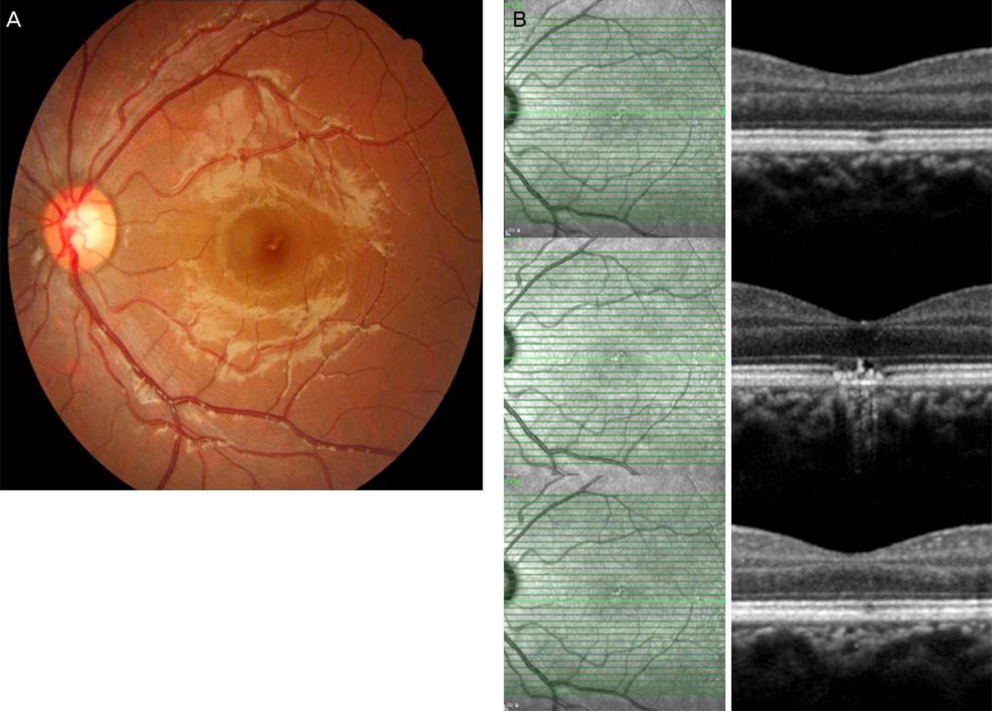
Figure 3. Fundus photograph (A) and SD-OCT (B) after 1 month of treatment show external limiting membrane was fully restored, and inner segment/outer segment line was partially restored, but the retinal scar at the foveal area, and retinal pigment epithelial complex injury remain in place. SD-OCT = spectral domain optical coherence tomography.
Reference
-
References
1. Choi SW, Chun KI, Lee SJ, Rah SH. A case of photic retinal injury associated with exposure to plasma arc welding. Korean J Ophthalmol. 2006; 20:250–3.
Article2. Ryan SJ. Retina. 5th ed.Vol. 2. Los Angeles: Elsevier Mosby;2013. p. 1555–8.3. Jeong WD, Hwang YH, Kim JS, Lee JH. Maculopathy from red laser pointer. J Korean Ophthalmol Soc. 2007; 48:1007–11.4. Robertson DM, McLaren JW, Salomao DR, Link TP. Retinopathy from a green laser pointer: a clinicopathologic study. Arch Ophthalmol. 2005; 123:629–33.5. Kim Martha, Kwon JW, Han YK. A case of green laser pointer injury to the macula. J Korean Ophthalmol Soc. 2008; 49:681–4.
Article6. Raoof N, Chan TK, Rogers NK, et al. ‘Toy’ laser macular burns in children. Eye (Lond). 2014; 28:231–4.
Article7. Barkana Y, Belkin M. Laser eye injuries. Surv Ophthalmology. 2000; 44:459–78.
Article8. Schulmeister K, Jean M. The risk of retinal injury from Class 2 and visible Class 3R lasers, including medical laser aiming beams. Medical Laser Application. 2010; 25:99–110.
Article9. Ham WT Jr, Geeraets WJ, Mueller HA, et al. Retinal burn thresh-olds for the helium-neon laser in the rhesus monkey. Arch Ophthalmol. 1970; 84:797–809.
Article10. Robertson DM, McLaren JW, Salomao DR, Link TP. Retinopathy from a green laser pointer: a clinicopathologic study. Arch Ophthalmol. 2005; 123:629–33.11. Sell CH, Bryan JS. Maculopathy from handheld diode laser pointer. Arch Ophthalmol. 1999; 117:1557–8.
Article12. Lam TT, Takahashi K, Fu J, Tso MO. Methylprednisolone therapy in laser injury of the retina. Graefes Arch Clin Exp Ophthalmol. 1993; 231:729–36.
Article13. Rosner M, Solberg Y, Turetz J, Belkin M. Neuroprotective therapy for argon-laser induced retinal injury. Exp Eye Res. 1997; 65:485–95.
Article14. Hirsch DR, Booth DG, Schocket S, Sliney DH. Recovery from pulsed-dye laser retinal injury. Arch Ophthalmol. 1992; 110:1688–9.
Article15. Zwick H, Stuck BE, Dunlap W, et al. Accidental bilateral Q-switch-ed neodymium laser exposure: treatment and recovery of visual function. In BiOS'98 International Biomedical Optics Symposium. International Society for Optics and Photonics. 1998; 3254:80–89.16. Cai YS, Xu D, Mo X. Clinical, pathological and photochemical studies of laser injury of the retina. Health Phys. 1989; 56:643–6.17. Lam TT, Tso MO. Retinal injury by neodymium: YAG laser. Retina. 1996; 16:42–6.18. Liu HF, Gao GH, Wu DC, et al. Ocular injuries from accidental laser exposure. Health Phys. 1989; 56:711–6.
- Full Text Links
-
- Actions
-
Cited
- CITED
-
- Close
- Share
-
- Similar articles
-
- Maculopathy from Red Laser Pointer
- A Case of Green Laser Pointer Injury to the Macula
- Novel Adjuvant Method to Assist Localisation of a Cyclodialysis Cleft
- A Case of Green Laser Pointer-induced Atypical Impending Macular Hole Treated with Vitrectomy in a Pediatric Patient
- Clinical Analysis on Focal Laser Treatment of Diabetic Maculopathy
