J Korean Ophthalmol Soc.
2014 Jul;55(7):984-990.
Comparative Analysis of Corneal Refractive Power Measured with AL-Scan(R), Autokeratometer, and Pentacam(R)
- Affiliations
-
- 1Department of Ophthalmology, Busan Paik Hospital, Inje University College of Medicine, Busan, Korea.
- 2Shinsegae Eye Clinic, Busan, Korea. happytriad@gmail.com
Abstract
- PURPOSE
To investigate clinical availability of AL-Scan(TM) (Nidek, Gamagori, Japan) by comparing corneal refractive power with AL-Scan(TM), Autokeratometer(TM) (Topcon KR-1, Tokyo, Japan) and Pentacam(TM) (Oculus, Wetzlar, Germany) devices.
METHODS
Seventy-one patients (142 eyes) who visited our hospital for refractive surgery were tested using AL-Scan(R), Autokeratometer and Pentacam(R) and corneal refractive power was compared among devices.
RESULTS
When comparing measurements with AL-Scan(R), Autokeratometer and Pentacam(R), the mean corneal refractive power was 43.37 +/- 1.32 D (2.4 mm zone), 43.35 +/- 1.32 D (3.3 mm zone), 43.36 +/- 1.35 D, and 43.35 +/- 1.36 D respectively and showed no significant differences. Corneal refractive power had strongly positive linear correlation (p < 0.001) and Bland-Altman plots showed high degree of agreement among AL-Scan(R), Autokeratometer and Pentacam(R) devices.
CONCLUSIONS
Because measuring ocular biometry with AL-Scan(R) including axial length, intraocular lens power calculation and topography simultaneously is possible, clinical use is convenient. Corneal refractive power was not different when compared with autokeratometer and Pentacam(R) devices, thus, AL-Scan(R) can be used in the clinical environment.
Figure
-

Figure 1. Pearson correlation of corneal refractive power (K) measured with AL-Scan®, Autokeratometer and Pentacam®.
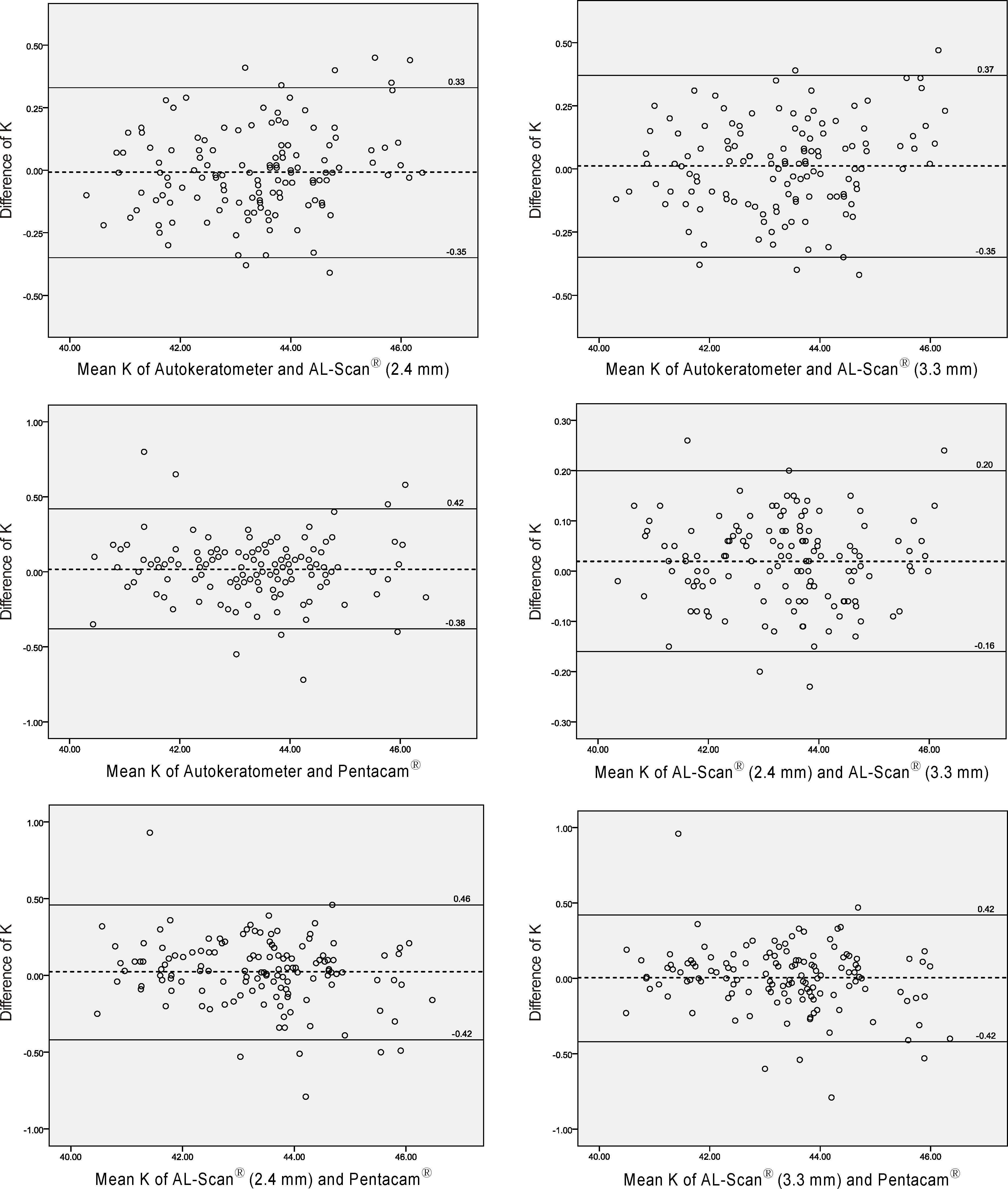
Figure 2. Bland-Altman plots of corneal refractive power (K) measured with AL-Scan®, Autokeratometer and Pentacam®.
Reference
-
References
1. Koranyi G, Lydahl E, Norrby S, Taube M. Anterior chamber depth measurement: a-scan versus optical methods. J Cataract Refract Surg. 2002; 28:243–7.
Article2. Maeng HS, Ryu EH, Chung TY, Chung ES. Effects of anterior chamber depth and axial length on refractive error after intraocular lens implantation. J Korean Ophthalmol Soc. 2010; 51:195–202.
Article3. Norrby S. Sources of error in intraocular lens power calculation. J Cataract Refract Surg. 2008; 34:368–76.
Article4. Olsen T. Prediction of the effective postoperative (intraocular lens) anterior chamber depth. J Cataract Refract Surg. 2006; 32:419–24.
Article5. Qazi MA, Cua IY, Roberts CJ, Pepose JS. Determining corneal power using Orbscan II videokeratography for intraocular lens calculation after excimer laser surgery for myopia. J Cataract Refract Surg. 2007; 33:21–30.
Article6. Rufer F, Schroder A, Arvani MK, Erb C. Central and peripheral corneal pachymetry–standard evaluation with the Pentacam system. Klin Monbl Augenheilkd. 2005; 222:117–22.7. Lee DM, Ahn JM, Seo KY, et al. Comparison of corneal measurement values between two types of topography. J Korean Ophthalmol Soc. 2012; 53:1584–90.
Article8. Kim SI, Kang SJ, Oh TH, et al. Accuracy of ocular biometry and postoperative refraction in cataract patients with AL-Scan(R). J Korean Ophthalmol Soc. 2013; 54:1688–93.9. Holladay JT, Prager TC, Ruiz RS, et al. Improving the predictability of intraocular lens power calculations. Arch Ophthalmol. 1986; 104:539–41.
Article10. Mamalis N. Complications of foldable intraocular lenses requiring explanation or secondary intervention–1998 survey. J Cataract Refract Surg. 2000; 26:766–72.11. Jo DH, Oh JY, Kim MK, et al. Corneal power estimation using Orbscan II videokeratography in eyes with previous corneal refractive surgeries. J Korean Ophthalmol Soc. 2009; 50:1730–4.
Article12. Findl O, Drexler W, Menapace R, et al. Improved prediction of intraocular lens power using partial coherence interferometry. J Cataract Refract Surg. 2001; 27:861–7.
Article13. Speicher L. Intra-ocular lens calculation status after corneal refractive surgery. Curr Opin Ophthalmol. 2001; 12:17–29.
Article14. Rosa N, Lanza M, Borrelli M, et al. Comparison of central corneal thickness measured with Orbscan and Pentacam. J Refract Surg. 2007; 23:895–9.
Article15. Butcher JM, O'Brien C. The reproducibility of biometry and keratometry measurements. Eye. 1991; 5:708–11.
Article16. Karabatsas CH, Cook SD, Papaefthymiou J, et al. Clinical evaluation of keratometry and computerised videokeratography: intraobserver and interobserver variability on normal and astigmatic corneas. Br J Ophthalmol. 1998; 82:637–42.
Article17. Han JM, Choi HJ, Kim MK, et al. Comparative analysis of corneal refraction and astigmatism measured with autokeratometer, IOL Master, and topography. J Korean Ophthalmol Soc. 2011; 52:1427–33.
Article18. Kim S, Chung SK. Comparison of corneal curvatures obtained with different devices. J Korean Ophthalmol. 2012; 53:618–25.
Article19. Savini G, Carbonelli M, Sbreglia A, et al. Comparison of anterior segment measurements by 3 Scheimpflug tomographers and 1 Placido corneal topographer. J Cataract Refract Surg. 2011; 37:1679–85.
Article20. Kawamorita T, Uozato H, Kamiya K, et al. Repeatability, reproducibility, and agreement characteristics of rotating Scheimpflug photography and scanning-slit corneal topography for corneal power measurement. J Cataract Refract Surg. 2009; 35:127–33.
Article21. Jain R, Dilraj G, Grewal SP. Repeatability of corneal parameters with Pentacam after laser in situ keratomileusis. Indian J Ophthalmol. 2007; 55:341–7.
- Full Text Links
-
- Actions
-
Cited
- CITED
-
- Close
- Share
-
- Similar articles
-
- Measurement Comparison of Anterior Segment Parameters between AL-Scan(R) and Pentacam(R)
- Comparison of Anterior Segment Measurements between Dual and Single Scheimpflug Camera
- Comparative Analysis of Corneal Refraction and Astigmatism Measured with Autokeratometer, IOL Master, and Topography
- Correlations among Refractive Error, Axial Length, and Corneal Power in Childhood
- Clinical Reliability of the Topolyzer Vario Instrument for Measurement of Corneal Refractive Power
