A Case of MALT Lymphoma with Left Inferior Rectus Muscle Invasion
- Affiliations
-
- 1Department of Ophthalmology, Soonchunhyang University Cheonan Hospital, Soonchunhyang University College of Medicine, Cheonan, Korea. ophdrkim@schmc.ac.kr
Abstract
- PURPOSE
To report an unusual case of mucosa-associated lymphoid tissue (MALT) lymphoma localized to the left inferior rectus muscle.
CASE SUMMARY
A 52-year-old male presented with double vision 6 months in duration, which was exacerbated in the down-gaze. On initial examination, 6 and 30 prism diopters (PDs) of left hypertropia were observed in primary gaze and down gaze, respectively. Prominently limited infraduction was also observed in his left eye. Computed tomography (CT) revealed contrast enhancing mass on the left inferior rectus muscle. He was diagnosed with suspicious orbital pseudotumor and treated with systemic steroid for 2 months. Double vision and limited infraduction was improved and the mass size was decreased on CT. After 21 months, the patient revisited the clinic with the same symptoms. In the down-gaze, 35 PDs of hypertropia were observed in his left eye along with limited infraduction. CT revealed an enlarged mass and left superior rectus muscle recession along with an incisional biopsy of the left inferior rectus muscle were performed. Infiltration by small lymphoid cells was detected with hematoxylin-eosin and immunohistochemical stainings. Subsequently, the patient was diagnosed with MALT lymphoma localized to the left inferior rectus muscle.
CONCLUSIONS
This is the first description of MALT lymphoma with inferior rectus muscle invasion in a Korean patient. In a patient with strabismus and limited duction, the possibility of extraocular muscle tumor including lymphoma should be considered. Homogenously contrast-enhanced mass on orbit CT can help in to make a diagnosing extraocular muscle lymphoma.
Keyword
MeSH Terms
Figure
-

Figure 1. Initial computed tomography revealed 4.0 × 2.0 cm sized homogenously enhancing mass which involving left inferior rectus muscle. (A) Axial view. (B) Coronal view.
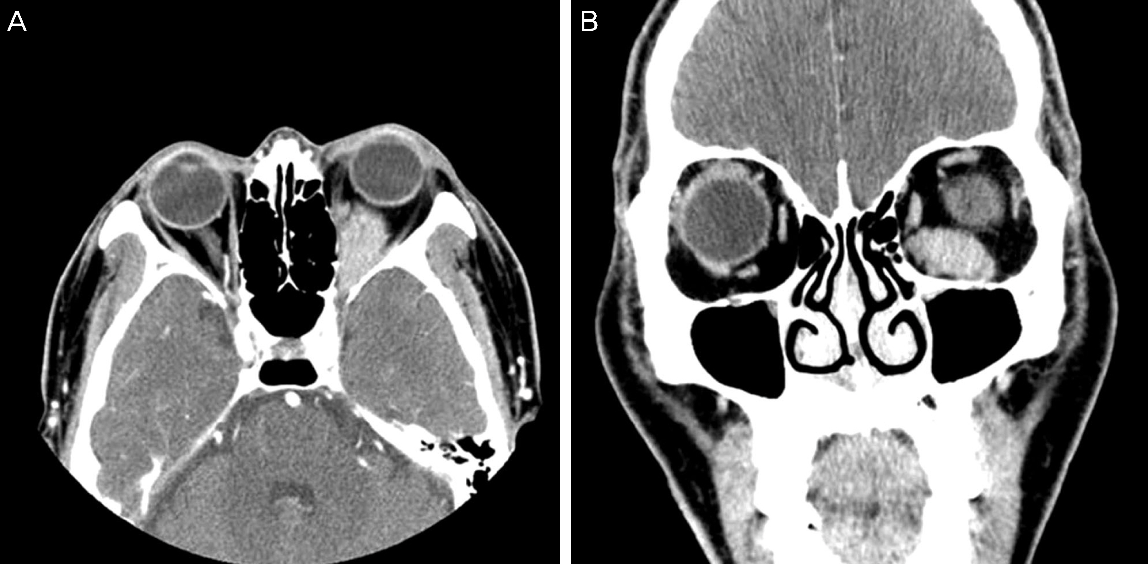
Figure 2. Preoperative computed tomograph revealed 2.4 × 2.2 × 2.9 cm sized homogenously enhancing mass which involving left inferior rectus muscle. (A) Axial view. (B) Coronal view.
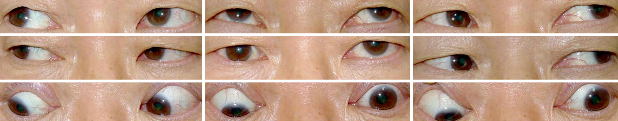
Figure 3. Preoperative external 9 gaze photograph showing left hypertropia in primary gaze and down gaze along with limited in-fraduction in the left eye.
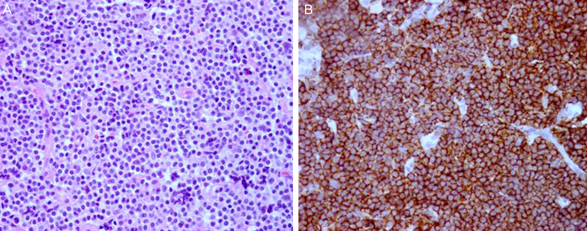
Figure 4. Results of Hematoxylin-Eosin staining (H-E) and Immunohistochemical staining (IHC). (A) The cells appear moderate amounts of pale cytoplasm, irregular nuclear contours, and dispersed chromatin resembling monocytoid B-cells (H-E, × 400). (B) The neoplastic cells express diffusely and strongly the B-cell marker CD20 (IHC, × 400).
Reference
-
References
1. Keleit D, Flickinger JC, Hobson SR, Mittal BB. Radiotherapy of lymphoproliferative disease of the orbit. Am J Clin Oncol. 1992; 15:422–7.2. Freeman C, Berg JW, Cutler SJ. Occurrence and prognosis of ex-tranodal lymphomas. Cancer. 1972; 29:252–60.
Article3. Cho EY, Han JJ, Ree HJ, et al. Clinicopathologic analysis of ocular adnexal lymphomas: extranodal marginal zone b-cell lymphoma constitutes the vast majority of ocular lymphomas among Koreans and affects younger patients. Am J Hematol. 2003; 73:87–96.
Article4. Ferry JA, Fung CY, Zukerberg L, et al. Lymphoma of the ocular ad-nexa: A study of 353 cases. Am J Surg Pathol. 2007; 31:170–84.
Article5. Abalo-Lojo JM, Baleato-Gonzalez S, Abdulkader I, Gonzalez F. Extraocular muscle involvement in MALT lymphomas. Orbit. 2011; 30:186–8.
Article6. Izambart C, Robert PY, Petellat F, et al. Extraocular muscle in-volvement in marginal zone B-cell lymphomas of the orbit. Orbit. 2008; 27:345–9.
Article7. Liesegang TJ. Ocular adnexal lymphoproliferative lesions. Mayo Clin Proc. 1993; 68:1003–10.
Article8. Lee SJ, Jung JH, Choi HY. Analysis of clinical features and prog-nostic factor analysis of orbital and adnexal lymphoma. J Korean Ophthalmol Soc. 2013; 54:12–8.
Article
- Full Text Links
-
- Actions
-
Cited
- CITED
-
- Close
- Share
-
- Similar articles
-
- Resection and Transposition of the Inferior Oblique for Hypertropia due to the Inferior Rectus Loss
- Malignant Lymphoma Occurred in Lateral Rectus Muscle
- A Case of Malignant Lymphoma of Lateral Rectus Muscle
- The Effect of Anteriorization of The Inferior Oblique Muscle in +3 or +4 Inferior Oblique Overaction
- The Effect of Modified Anterior Transposition of the Inferior Oblique Muscle for Hypertropia in Superior Oblique Muscle Palsy with Inferior Oblique Muscle Overaction
