J Korean Ophthalmol Soc.
2012 Oct;53(10):1488-1492.
Trypan Blue-Assisted Measurements of Anatomical Positions of the Superior Rectus Muscle and Superior Oblique Muscle in Enucleated Eyes
- Affiliations
-
- 1Department of Ophthalmology, Kyungpook National University School of Medicine, Daegu, Korea. byjun424@hotmail.com
Abstract
- PURPOSE
To recognize the anatomical positions of the superior oblique muscle in enucleated eyes using trypan blue.
METHODS
Twenty-two surgically-enucleated eyes of 11 bodies were studied. The shortest distance from the nasal insertion of superior rectus to the anterior end of the superior oblique tendon, the distance from the temporal insertion of superior rectus to the anterior end of the superior oblique insertion, and the greatest width of superior oblique tendon insertion were measured by caliper 3 consecutive times. The average values in each of the above 3 points were calculated, and values prior to and after trypan blue staining were compared.
RESULTS
Prior to staining with trypan blue, the average distance from the nasal insertion of superior rectus to the anterior end of the superior oblique tendon was 4.97 mm and the average distance from the temporal insertion of superior rectus to the anterior end of the superior oblique insertion was 7.57 mm; after staining with trypan blue, the average values were 5.09 mm and 7.65 mm, respectively. There was no statistically meaningful difference in values prior to and after staining (p > 0.05). Prior to staining, the average value of the greatest width of the superior oblique tendon was 10.32 mm, and after staining with trypan blue, the average value increased to 10.76 mm. There was a statistically meaningful difference between the values (p = 0.02).
CONCLUSIONS
Trypan blue staining helped to recognize the location and the width of the superior oblique tendon more precisely.
Keyword
Figure
-
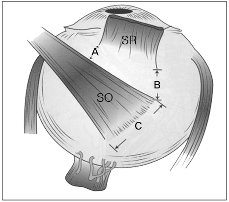
Figure 1 Measurements of anatomical positions. (A) The distance from the nasal insertion of the superior rectus (SR) to the anterior end of the superior oblique tendon (SO), (B) The distance from the temporal insertion of the superior rectus (SR) to the anterior end of the superior oblique insertion (SO), (C) The greatest width of the superior oblique tendon (SO) insertion.

Figure 2 (A) After enucleation, dissection around the superior rectus muscle (SR) and superior oblique muscle (SO), (B) Application of trypan blue at the tendon of the superior oblique muscle.
Reference
-
1. Lee JB. Current Concepts in Strabismus. 2004. 1st ed. Seoul: Naewae Haksool;7–8.2. Saxena R, Sinha A, Sethi H, Menon V. Trypan blue-assisted posterior tenectomy of the superior oblique. J Pediatr Ophthalmol Strabismus. 2007. 44:45–46.3. Parks MM, Helveston EM. Direct visualization of the superior oblique tendon. Arch Ophthalmol. 1970. 84:491–494.4. Lozano MJ, Santiago AP, Rosenbaum AL. Rosenbaum AL, Santiago AP, editors. Superior oblique procedures. Clinical Strabismus Management Principles and Surgical Techniques. 1999. Philadelphia: WB Saunders;463–464.5. Fuchs E. Beiträge zur normalen Anatomie des Augapfels. Graefes Arch Clin Exp Ophthalmol. 1884. 30:1–60.6. Apt L. An anatomical reevaluation of rectus muscle insertions. Trans Am Ophthalmol Soc. 1980. 78:365–375.7. Paik HJ, Cho YA. Insertion of horizontal rectus muscles in strabismus. J Korean Ophthalmol Soc. 1989. 30:761–766.8. Lee YC, Yang SW. Anatomical evaluations of the location and insertion shape of horizontal rectus muscle. J Korean Ophthalmol Soc. 1995. 36:1357–1362.9. Shin HM, Lew H, Yun YS. Muscle width and distance from limbus to muscle insertion site in strabismus patient. J Korean Ophthalmol Soc. 2005. 46:1387–1392.10. Parks MM. Duane TD, editor. Extraocular muscles. Clinical Ophthalmology. 1987. v. 1:11th ed. Philadelphia: Haper and Row;chap. 1.11. Helveston EM. Atlas of Strabismus Surgery. 1985. 3rd ed. St. Louis: CV Mosby;56–57.12. von Noorden GK. Binocular Vision and Ocular Motility : Theory and Management of Strabismus. 1990. 4th ed. St. Louis: CV Mosby;44.13. Wobig JL. Ophthalmic Anatomy. 1981. 1st ed. San Francisco: American Academy of Ophthalmology;34.14. O'Sullivan E, Mitchell BS. An improved composition for embalming fluid to preserve cadavers for anatomy teaching in the United Kingdom. J Anat. 1993. 182(Pt 2):295–297.15. Ward SR, Lieber RL. Density and hydration of fresh and fixed human skeletal muscle. J Biomech. 2005. 38:2317–2320.16. Dada VK, Sharma N, Sudan R, et al. Anterior capsule staining for capsulorhexis in cases of white cataract: comparative clinical study. J Cataract Refract Surg. 2004. 30:326–333.17. Lee KL, Dean S, Guest S. A comparison of outcomes after indocyanine green and trypan blue assisted internal limiting membrane peeling during macular hole surgery. Br J Ophthalmol. 2005. 89:420–424.18. Ignacio TS, Nguyen TT, Sarayba MA, et al. A technique to harvest Descemet's membrane with viable endothelial cells for selective transplantation. Am J Ophthalmol. 2005. 139:325–330.
- Full Text Links
-
- Actions
-
Cited
- CITED
-
- Close
- Share
-
- Similar articles
-
- Anatomical Evaluation of Extraocular Muscles and Relationships Among Macula and Optic Nerve in Enucleated Eye
- Strabismus Surgery on Congenital Oculomotor Nerve Palsied Eye
- The Effect of Modified Anterior Transposition of the Inferior Oblique Muscle for Hypertropia in Superior Oblique Muscle Palsy with Inferior Oblique Muscle Overaction
- A Study on the Anatomical Position of the Inferior Oblique Muscle Insertion in Primary Inferior Oblique Overaction
- Surgical Correction of Exotropia due to Oculomotor Nerve Palsy
