A Case of Retinal Injury During a Laser Show
- Affiliations
-
- 1Department of Ophthalmology, Gyeongju Hospital, Dongguk University College of Medicine, Gyeongju, Korea. meinkamf@hanmir.com
Abstract
- PURPOSE
To report a case of macular injury after exposure to a high energy laser beam used in a laser show. CASE: A 19-year-old female presented 2 days after exposure to a high energy laser beam at a laser show in a night club with decreased vision in her right eye. The patient's best corrected visual acuity of the right eye was hand motion. Fundus examination reveald a retinal swelling in the macular area approximately 5 disc diameter in size and a submacular hemorrhage. Fluorescein angiography of the right eye showed marked hypofluorescence in the macular area and optical coherence tomography (OCT) showed a neurosensory retinal detachment with a macular edema. Three years after exposure, the visual acuity of the right eye improved to 20/600. The fundus revealed scar and depigmented area at the macula.
CONCLUSIONS
High-energy laser devices at laser shows should be used carefully with safety education and strict control and can provoke irreversible eye damage if not managed adequately.
Keyword
MeSH Terms
Figure
-
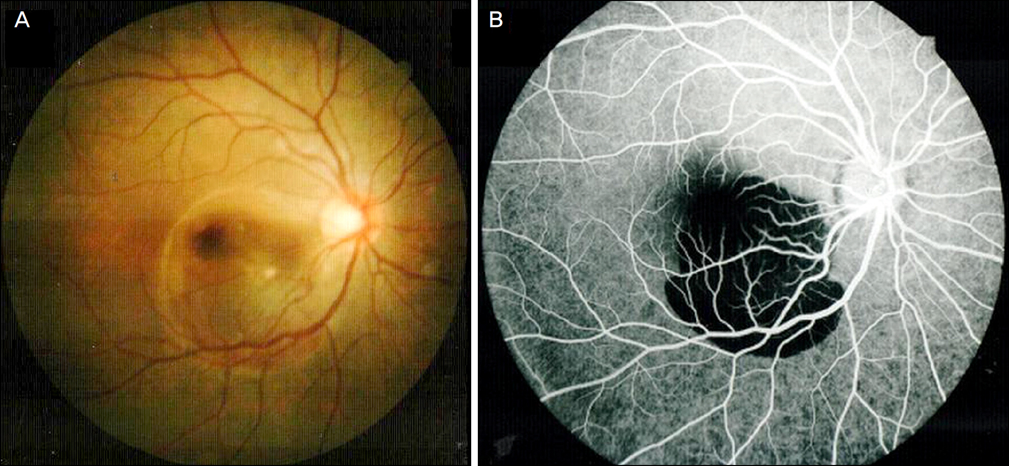
Figure 1. (A) Initial fundus photograph of the right eye, two days after exposure showed about a five disc diameter sized retinal swelling at the macula and a submacular hemorrhage. (B) Initial fluorescein angiograph of the right eye, two days after exposure to laser beam showed a marked hypofluorescence area at the macula.
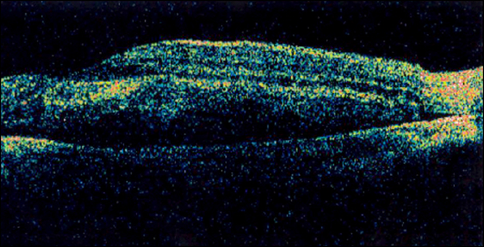
Figure 2. Optical coherence tomograph (OCT) of the right eye, two days after exposure to laser beam. There was neurosensory retinal detachment with a macular edema.
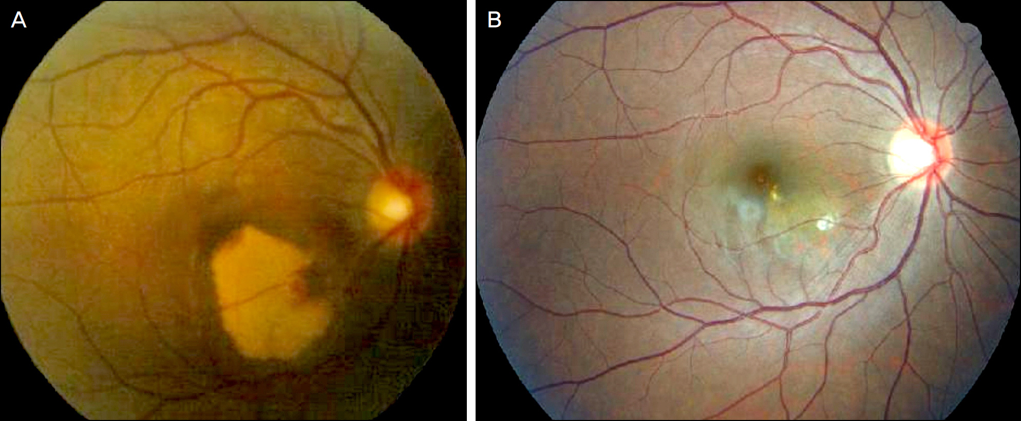
Figure 3. Fundus photographs (A) One month after exposure, fundus photography revealed subretinal exudates and macular edema. (B) Three years later, fundus photography revealed a rounded, slightly irregular depigmented area with well defined margin and scar at the macula.
Reference
-
References
1. Barkana Y, Belkin M. Laser eye injuries. Surv Ophthalmol. 2000; 44:459–78.
Article2. Cai YS, Xu D, Mo X. Clinical, pathological and photochemical studies of laser injury of the retina. Health Phys. 1989; 56:643–6.3. Wolfe JA. Laser retinal injury. Mil Med. 1985; 150:177–85.
Article4. Gabel VP, Birngruber R, Lorenz B, Lang GK. Clinical observations of six cases of laser injury to the eye. Health Phys. 1989; 56:705–10.
Article5. Mainster MA. Blinded by the light–not! Arch Ophthalmol. 1999; 117:1547–8.
Article6. Mainster MA, Timberlake GT, Warren KA, Sliney DH. Pointers on laser pointers. Ophthalmology. 1997; 104:1213–4.
Article7. Marshall J. The safety of laser pointers: myths and realities. Br J Ophthalmol. 1998; 82:1335–8.
Article8. Yolton RL, Citek K, Schmeisser E, et al. Laser pointers: toys, nui-sances, or significant eye hazards? J Am Optom Assoc. 1999; 70:285–9.9. Zamir E, Chowers I. Concerns about laser pointers and macular damage. Arch Ophthalmol. 2001; 119:1731–2.10. Jeong WD, Hwang YH, Kim JS, Lee JH. Maculopathy from red laser pointer. J Korean Ophthalmol Soc. 2007; 48:1007–11.11. Kim M, Kwon JW, Han YK. A case of green laser pointer injury to the macula. J Korean Ophthalmol. 2008; 49:681–4.
Article12. Ryan S. Photic Retinal Injury and Safety. RETINA. 3th ed.2. Los Angeles: Elsever Mosby;2001. p. 1797–805.13. Alhalel A, Glovinsky Y, Treister G, et al. Long-term follow up of accidental parafoveal laser burns. Retina. 1993; 13:152–4.
Article14. Dennis JE. Amendments to the Center for Devices and Radiological Health federal performance standard for laser products. J Laser Appl. 1997; 9:301–5.
Article
- Full Text Links
-
- Actions
-
Cited
- CITED
-
- Close
- Share
-
- Similar articles
-
- A Case of Retinal Injury by Neodymium: YAG Laser
- Laser Photocoaculation Treatment in a Case of Circumscribged Choroidal hmangioma Associated with Serous Retinal Detachment
- Laser Photocoagulation Repair of Recurrent Macula-Sparing Retinal Detachments
- Drainage of Subretinal Fluid with the Diode Laser
- The Characteristics of Non-Retinal Lesions in the Ultra-Wide Field Scanning Laser Ophthalmoscope Image
