Recurrent Alveolar Soft-Part Sarcoma With Concurrent Involvement of the Orbit and Multiple Sites of the Body
- Affiliations
-
- 1Department of Ophthalmology, The Catholic University of Korea School of Medicine, Seoul, Korea. laty@catholic.ac.kr
Abstract
- PURPOSE
To report a rare case of recurrent alveolar soft-part sarcoma (ASPS) with concurrent involvement of the orbit and multiple sites of the body that was removed successfully by surgery.
CASE SUMMARY
A 37-year-old woman presented with diplopia at the left lateral gaze and proptosis of the left eye. Two years earlier, the patient had a mass excision of the right gluteus maximus and the left orbit, and ASPS was histopathologically diagnosed at that time. In addition, the patient had been treated with chemotherapy and radiation therapy. On radiologic examination, recurrent tumor of the left orbit was found and surgically removed. The histopathologic examination showed that tumor cells were arranged in an alveolar pattern divided by fibrous septa and contained abundant granules in the cytoplasm, typically consistent with ASPS. Postoperatively, the symptoms of diplopia and proptosis improved.
CONCLUSIONS
ASPS can occur and recur in the orbit as well as systemically. In addition, at the time of surgical removal of ASPS in the orbit, the surgeon should be particularly cautious of massive bleeding.
Keyword
MeSH Terms
Figure
-

Figure 1. (A) Pelvic MRI shows a large lobulated hypervascular soft tissue mass in the right gluteus maximus muscle about 11.8×9.3 cm in diameter. (B) Brain CT shows bone destructions and formations of the left orbit and sphenoid bone with contrast enhancing of a soft tissue mass (arrow) in the left orbit.
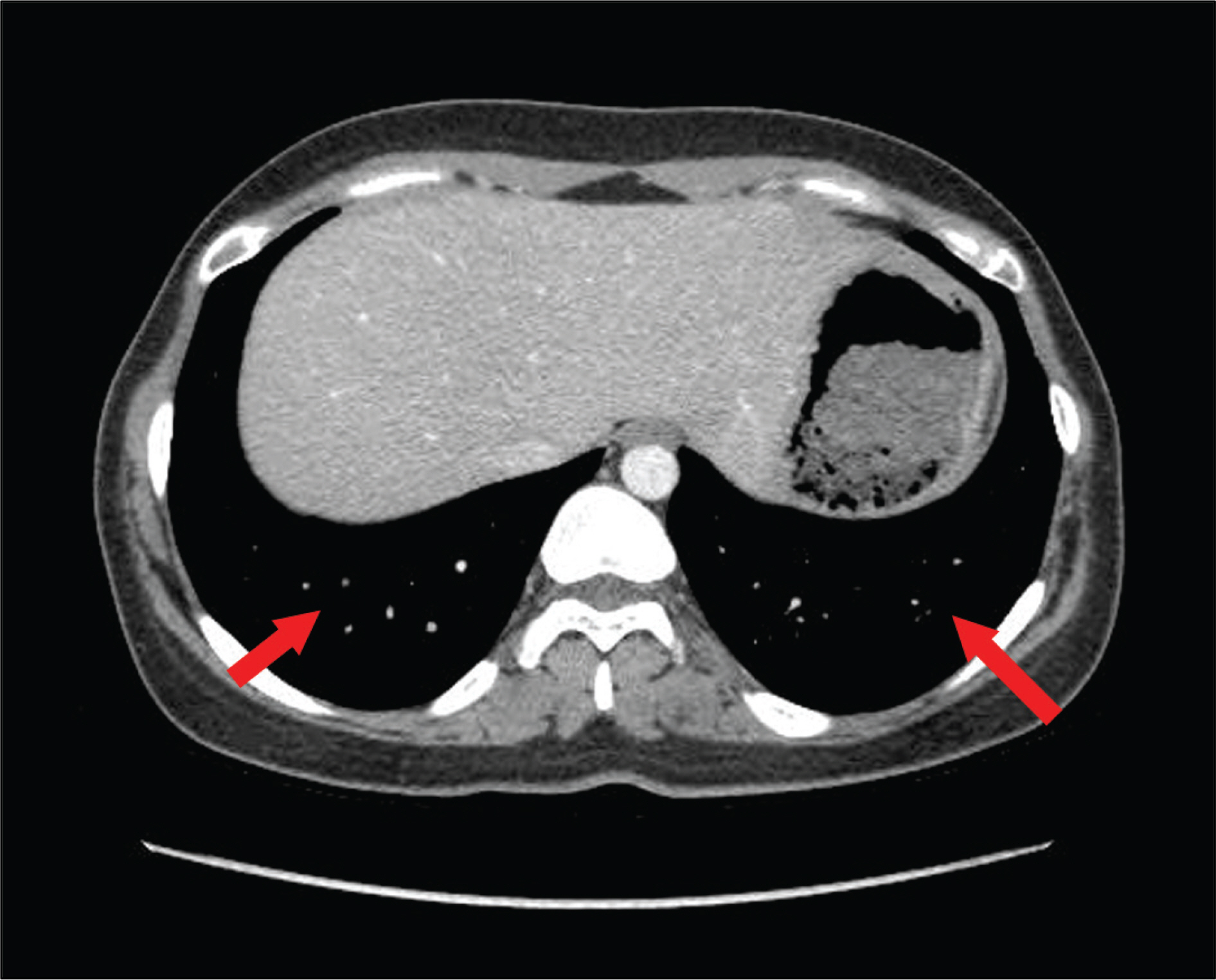
Figure 2. Chest CT shows multiple small nodules (arrows) in both lungs.
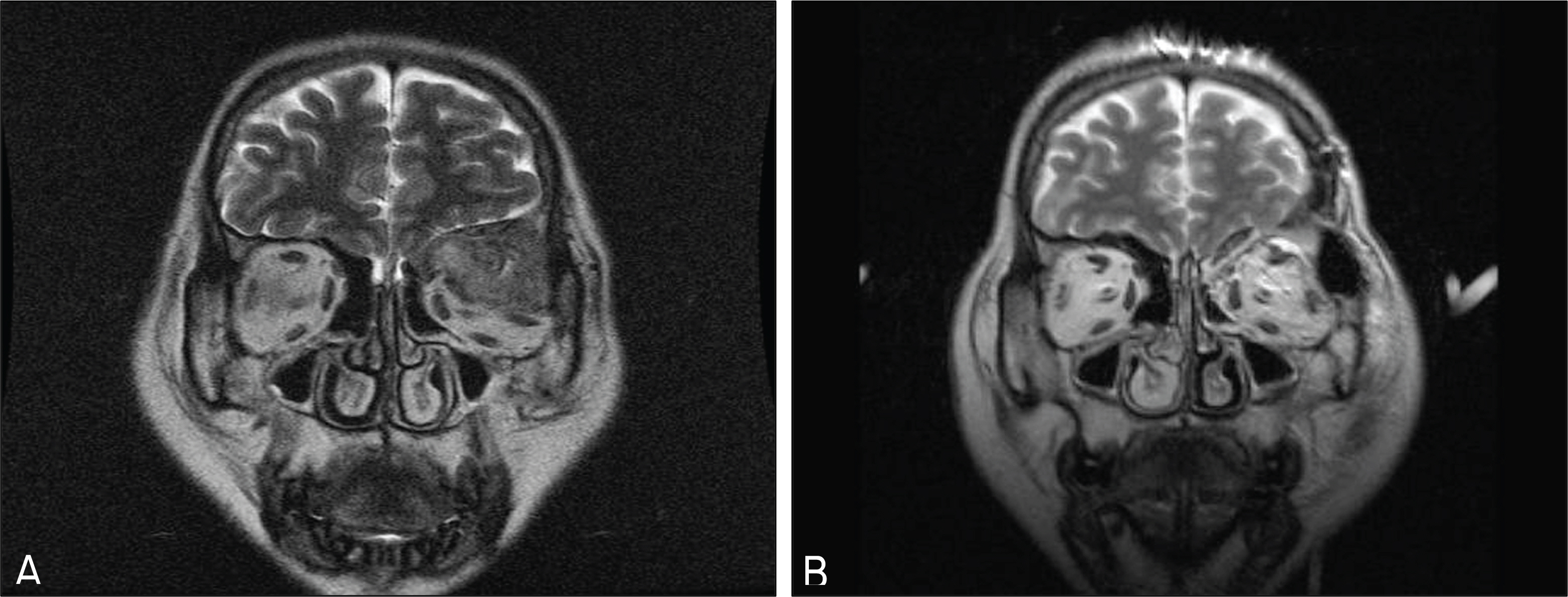
Figure 3. (A) Preoperative brain MRI shows left orbital roof destruction with intraorbital invasion of a soft tissue mass. (B) Post-operative brain MRI shows that the part of the left Laserassistedlateral orbital wall, and intraorbital mass were removed.
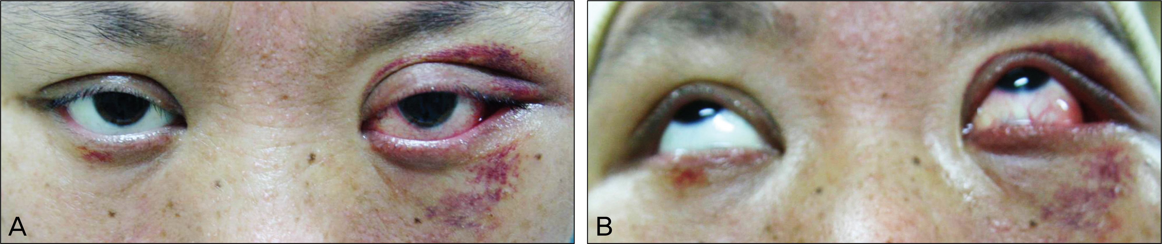
Figure 4. Patient shows proptosis of the left eye with periorbital ecchymosis and conjunctival injection (A: Frontal view, B: Chin up view).
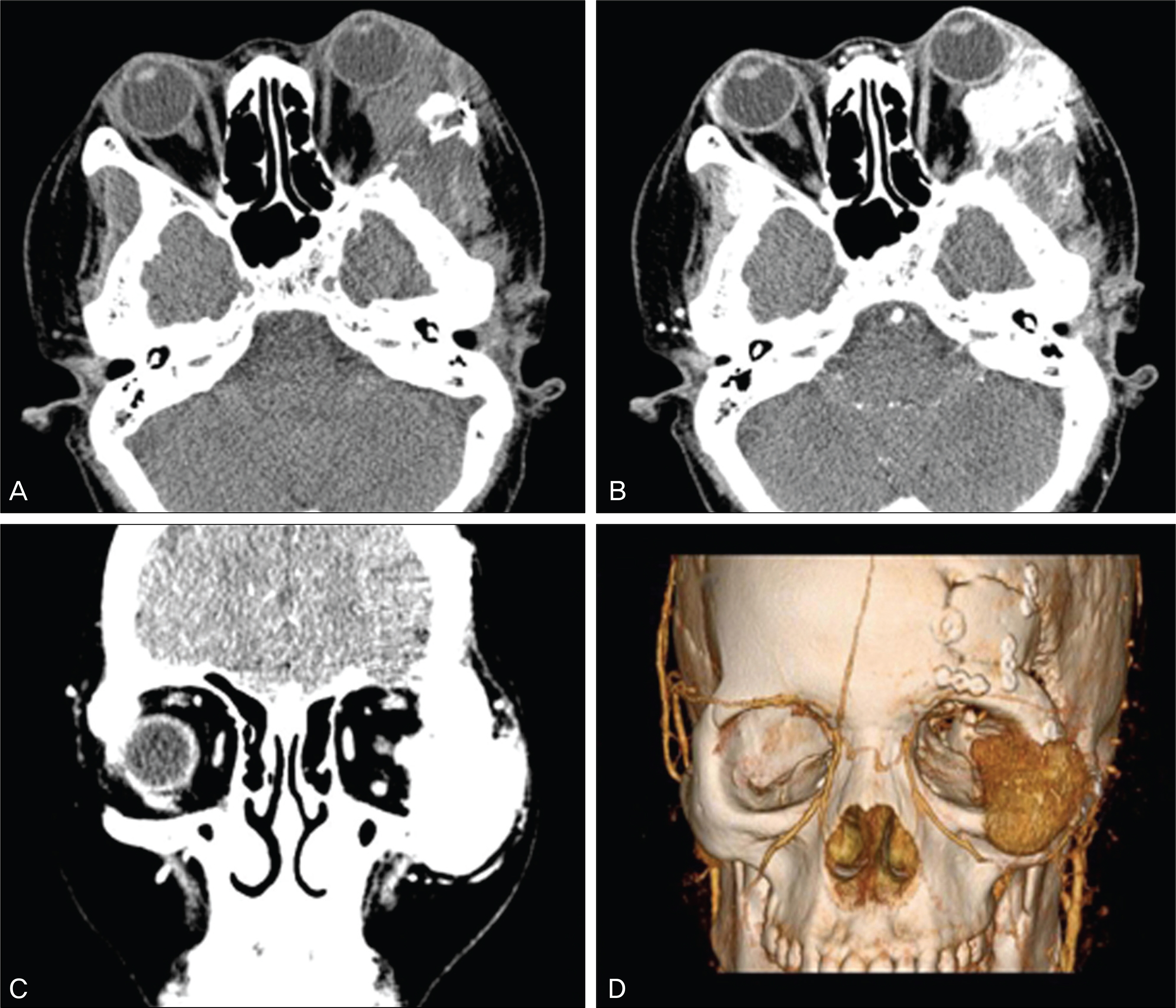
Figure 5. Preoperative facial CT and 3 dimensional CT shows well enhanced soft tissue mass in the left orbit and zygomatic bone area. CT also shows mass invasion adjacent to the orbital apex and unclear outline of the left lateral rectus muscle. (A: Transverse section without contrast enhancement (CE), B: Transverse section with CE, C: Coronal section with CE, D: 3 dimensional view).

Figure 6. (A) Through lateral canthotomy and conjunctival incision, a dark red-colored, capsulated soft tissue mass (arrow) was found underneath. (B) An almost totally exposed mass is seen. (C) Removed mass was 4×3×2.5 cm in size. (D) Photograph of post-operative 1 month shows improved proptosis.

Figure 7. The tumor cells were arranged in an alveolar pattern divided by fibrous septa (A, H & E stain, ×100) and contained abundant granular cytoplasm (B, H & E stain, ×400). PAS positive intracytoplasmic crystal-like granules were observed character-istically (C, PAS stain, ×400).
Reference
-
References
1. Christopherson WM, Foote FW Jr, Stewart FW. Alveolar soft part sarcoma. Structurally characteristic tumor of uncertain histogenesis. Cancer. 1952; 5:100–11.2. Spector RA, Travis LW, Smith J. Alveolar soft part sarcoma of the head and neck. Laryngoscope. 1979; 89:1301–6.
Article3. Ackerman LV. Sugical pathology. 3rd ed.St. Louis: C.V. Mosby Co.;1964. p. 992.4. Lee YM, Kim HC, Lee SH, et al. A case of alveolar soft part sarcoma in the orbit. J Korean Ophthalmol Soc. 1973; 14:35–9.5. Yang SE, Kim SM. A case of alveolar soft part sarcoma in the orbit. J Korean Ophthalmol Soc. 1980; 21:95–7.6. Lee DC, Lee SY, Chang MH, et al. A case of alveolar soft part sarcoma in the orbit diagnosed as cavernous hemangioma before operation. J Korean Ophthalmol Soc. 1999; 40:2299–303.7. Lieberman PH, Foote FW, Wtewart FW, et al. Alveolar Soft Part Sarcoma. JAMA. 1966; 198:1047–51.8. Ordonez NG. Alveolar soft part sarcoma: a review and update. Adv Anat Pathol. 1999; 6:125–39.9. Rubinstein MI, Drake AF, McClatchey KD, et al. Alveolar soft part sarcoma of the nasal cavity: Report of a case and a review of the literature. Laryngoscope. 1988; 98:1246–50.10. Enzinger FM, Weiss SW. Malignant soft tissue tumors of uncetain type. In soft tissue tumors. 3rd ed.St. Louis: Mosby Year Book;1995. p. 1067–74.11. Mukai M, Iri H, Nakajima T, et al. Alveolar soft part sarcoma. A review of the histogenesis and further studies based on electron microscopy, immunohistochemistry, and biochemistry. Am J Surg Pathol. 1983; 7:679–89.12. Philip H. Lieberman, Murray F. Brennan, Marek Kimmel. Alveolar soft part sarcoma, A clinicopathologic study of half a centry. Cancer. 1989; 63:1–13.13. Charles AP, Viet H, Shreyaskumar RP, et al. Alveolar soft part sarcoma, clinical course and patterns of metastasis in 70 patients treated at a single institution. Cancer. 2001; 91:585–91.14. Ahn SM, Oh JT, Choi SH. Surgical treatment for the alveolar soft part sarcoma. J Korean Surg Soc. 2004; 66:50–5.
