J Korean Ophthalmol Soc.
2010 Aug;51(8):1099-1106.
Bilateral Fundus Findings Using Examination Under Anesthesia in Patients Showing Vitreoretinopathy at Unilateral Posterior Pole
- Affiliations
-
- 1Department of Ophthalmology, Seoul National University College of Medicine, Seoul, Korea.
- 2Seoul Artificial Eye Center, Seoul National University Hospital Clinical Research Institute, Seoul, Korea.
Abstract
- PURPOSE
To study the fundus findings in the peripheral retina using examination under general anesthesia in patients showing vitreoretinopathy in the unilateral posterior pole of the eye.
METHODS
The records of 27 patients showing vitreoretinopathy in the unilateral posterior pole of the eye were retrospectively reviewed. Analysis of the records for the fundus finding was performed using indirect ophthlamoscopy at the outpatient clinic, fundus findings and results of fluorescein angiography during examination under general anesthesia.
RESULTS
The viteroretinopathies in the unilateral posterior poles were retinal vascular and vitreous abnormality including avascular area in seven eyes (16%), retinal dragging in 14 eyes (32%), retinal folding in 21 eyes (48%), and total tractional retinal detachment in two eyes (5%). The final clinical diagnoses were familial exudative vitreoretinopathy in 33 patients, incontinentia pigmenti in four eyes, and idiopathic vitreoretinopathy in seven eyes. Abnormal findings in the peripheral retina of the contralateral eye were observed in 27 eyes (61%) when examined under general anesthesia. Leakage in fluorescein angiography was found in 17 contralateral eyes (39%). The treatment including diode laser photocoagulation and cryotherapy was performed in eyes showing active leakage.
CONCLUSIONS
Examination of the bilateral fundus is important in patients showing vitreoretinopathy in the unilateral posterior pole. The examination under anesthesia was helpful for diagnosis, evaluation and treatment in patients showing vitreoretinopathy in the unilateral posterior pole of the eye.
Keyword
MeSH Terms
Figure
-
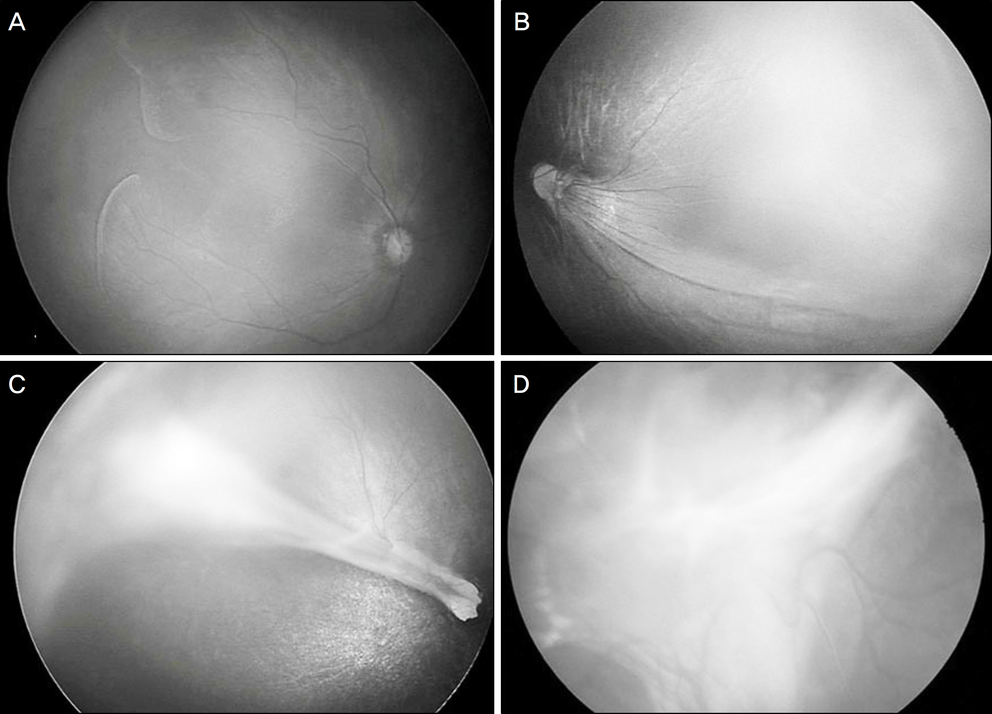
Figure 1. Vitereoretinopathy in unilateral posterior pole. (A) Retinal vascular abnormality, avascular area. (B) Retinal dragging. (C) Retinal fold. (D) Total tractional retinal detachment.

Figure 2. Fundus photographs and fluorescein angiographs of an 8-month-old girl. (A) Normal posterior pole in the right eye. (B) Retinal fold in the left eye. (C), (D), (E) Fluorescein angiographs show avascular areas in the temporal retina of the right eye (White arrow: the dropout of temporal vessels). (F) Fluorescein angiograph shows vascular leakage in the late phase (White arrowhead).
Reference
-
References
1. Multicenter trial of cryotherapy for retinopathy of prematurity. Snellen visual acuity and structural outcome at 5 1/2 years after randomization. Cryotherapy for Retinopathy of Prematurity Cooperative Group. Arch Ophthalmol. 1996; 114:417–24.2. Kim SY, Kim SJ, Yu YS. Early Laser Treatment of Rush-Type Retinopathy of Prematurity. J Korean Ophthalmol Soc. 2005; 46:448–55.3. Criswick VG, Schepens CL. Familial exudative vitreoretinopathy. Am J Ophthalmol. 1969; 68:578–94.
Article4. Canny CL, Oliver GL. Fluorescein angiographic findings in familial exudative vitreoretinopathy. Arch Ophthalmol. 1976; 94:1114–20.
Article5. Watzke RC, Stevens TS, Carney RG Jr. Retinal vascular changes of incontinentia pigmenti. Arch Ophthalmol. 1976; 94:743–6.
Article6. Francois J. Incontinentia pigmenti (Bloch-Sulzberger syndrome) and retinal changes. Br J Ophthalmol. 1984; 68:19–25.
Article7. Tasman W, Augsburger JJ, Shields JA, et al. Familial exudative vitreoretinopathy. Trans Am Ophthalmol Soc. 1981; 79:211–26.8. Benson WE. Familial exudative vitreoretinopathy. Trans Am Ophthalmol Soc. 1995; 93:473–521.9. Holmström G, Thorén K. Ocular manifestations of incontinentia pigmenti. Acta Ophthalmol Scand. 2000; 78:348–53.
Article10. Yu YS, Park KC. Retinal Vascular Changes and Treatment of incontinentia Pigmenti Eyes. J Korean Ophthalmol Soc. 1998; 39:1823–30.11. Shukla D, Singh J, Sudheer G, et al. Familial exudative vitreoretino- pathy (FEVR). Clinical profile and management. Indian J Opht- halmol. 2003; 51:323–8.12. Ebert EM, Mukai S. Familial exudative vitreoretinopathy. Int Ophthalmol Clin. 1993; 33:237–47.
Article13. Pendergast SD, Trese MT. Familial exudative vitreoretinopathy. Results of surgical management. Ophthalmology. 1998; 105:1015–23.
Article
