Tractional Retinal Detachment After Intravitreal Bevacizumab (Avastin(R)) Injection in Proliferative Diabetic Retinopathy
- Affiliations
-
- 1Department of Ophthalmology, Gachon University Gil Hospital, Incheon, Korea. eyedawns@gilhospital.com
Abstract
- PURPOSE
To report two cases of tractional retinal detachment after intravitreal bevacizumab injection.
CASE SUMMARY
(Case 1) A 48-year-old female with insulin-dependent diabetes mellitus and a high HbA1c level came to our clinic for fundus evaluation. The best corrected visual acuity (BCVA) was 1.0 in the right eye and funduscopic examination of the right eye revealed proliferative diabetic retinopathy with preretinal hemorrhage and a mild fibrovascular proliferative membrane around the optic disc. Intravitreal bevacizumab injection (1.25 mg) was performed before starting panretinal photocoagulation (PRP) to prevent macular edema after PRP. Three days after the injection, visual acuity decreased to 0.3 and funduscopic findings showed tractional retinal detachment. Vitrectomy was performed and visual acuity recovered to 1.0 four months after operation.
CONCLUSIONS
Intravitreal bevacizumab injection may cause tractional retinal detachment in poorly controlled insulin-dependent diabetes mellitus patients with fibrovascular proliferative membranes.
MeSH Terms
Figure
-
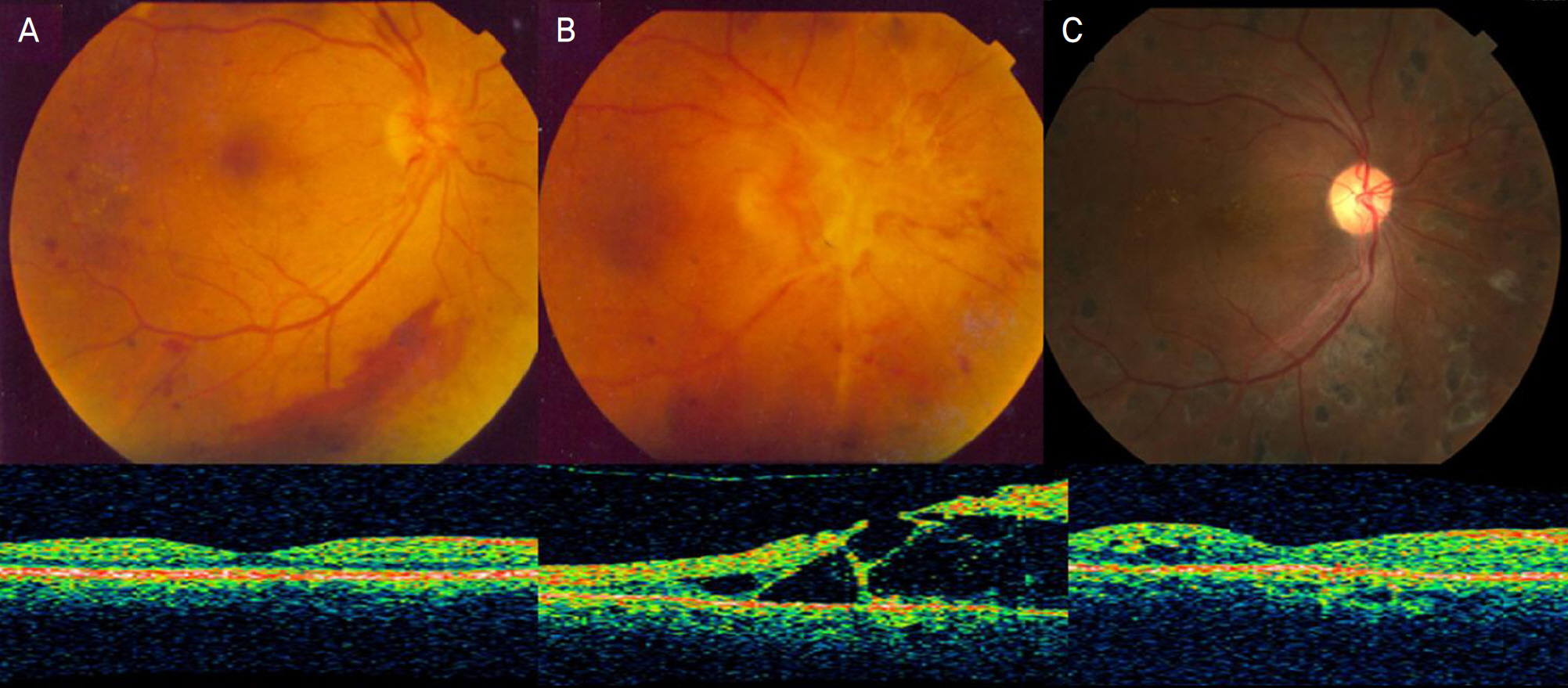
Figure 1. Fundus photography and optical coherence tomography of case 1 (A) At the initial visit, mild proliferative fibrovascular membrane and neovascularization around the optic disc and preretinal hemorrhage were seen in fundus photography. No macular edema was noted in OCT. (B) At one week after intravitreal bevacizumab injection, regressed but contractured fibrovascular proliferative membrane, tractional retinal detachment involving the fovea, decreased preretinal hemorrhage was seen in fundus photography. Tractional retinal detachment was noted in OCT. (C) At postoperative four months, well attached retina was seen in fundus photography and OCT.
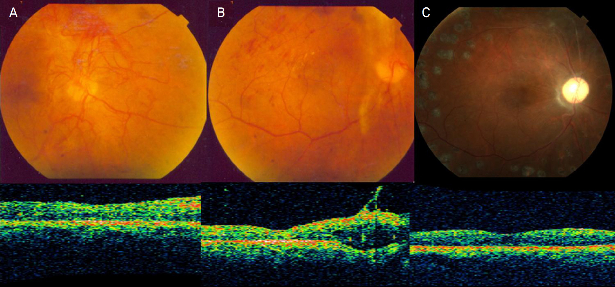
Figure 2. Fundus photography and optical coherence tomography of case 2 (A) At the initial visit, fibrovascular proliferative membrane around the optic disc, neovascularization at the disc and elsewhere, vitreous hemorrhage, macular edema were seen in fundus photography. No macular edema was noted in OCT. (B) At one month after intravitreal bevacizumab injection, dense fibrous tissue contraction, tractional retinal detachment, decreased vitreous hemorrhage were seen in fundus photography. Tractional retinal detachment was noted in OCT. (C) At three months after silicone oil removal, well attached retina was seen in fundus photography and OCT.
Reference
-
References
1. Ferrara N, Hillan KJ, Gerber HP, Novotny W. Discovery and development of bevacizumab, an anti-VEGF antibody for treating cancer. Nat Rev Drug Discov. 2004; 3:391–400.
Article2. Chen E, Park CH. Use of intravitreal bevacizumab as a preoperative adjunct for tractional retinal detachment repair in severe proliferative diabetic retinopathy. Retina. 2006; 26:699–700.
Article3. Yeoh J, Williams C, Allen P, et al. Avastin as an adjunct to vitrectomy in the management of severe proliferative diabetic retinopathy. Clin Experiment Ophthalmol. 2008; 36:449–54.4. Avery RL, Pearlman J, Pieramici DJ, et al. Intravitreal bevacizumab (Avastin) in the treatment of proliferative diabetic retinopathy. Ophthalmology. 2006; 113:1695–705.
Article5. Michels S, Rosenfeld PJ, Puliafito CA, et al. Systemic bevacizumab (Avastin) therapy for neovascular age-related macular degeneration twelve-week results of an uncontrolled open-label clinical study. Ophthalmology. 2005; 112:1035–47.6. Rosenfeld PJ, Schwartz SD, Blumenkranz MS, et al. Maximum tolerated dose of a humanized anti-vascular endothelial growth factor antibody fragment for treating neovascular age-related macular de-generation. Ophthalmology. 2005; 112:1048–53.
Article7. Mason JO 3rd, Yunker JJ, Vail R, McGwin G Jr. Intravitreal bevacizumab (Avastin) prevention of panretinal photocoagulation- induced complications in patients with severe proliferative diabetic retinopathy. Retina. 2008; 28:1319–24.8. Arevalo JF, Maia M, Flynn HW Jr, et al. Tractional retinal detach-ment following intravitreal bevacizumab (Avastin) in patients with severe proliferative diabetic retinopathy. Br J Ophthalmol. 2008; 92:213–6.
Article9. Jonas JB, Spandau UH, Schlichtenbrede F. Short-term complications of intravitreal injections of triamcinolone and bevacizumab. Eye. 2008; 22:590–1.
Article10. Mitamura Y, Ogata K, Oshitari T, et al. Retinal detachment with macular hole following intravitreal bevacizumab in patient with severe proliferative diabetic retinopathy. Br J Ophthalmol. 2008; 92:717–8.
Article11. Oshima Y, Shima C, Wakabayashi T, et al. Microincision vitrectomy surgery and intravitreal bevacizumab as a surgical adjunct to treat diabetic traction retinal detachment. Ophthalmology. 2009; 116:927–38.
Article
- Full Text Links
-
- Actions
-
Cited
- CITED
-
- Close
- Share
-
- Similar articles
-
- Effectiveness of Preoperative Intravitreal Bevacizumab Injections in Pars Plana Vitrectomy for Proliferative Diabetic Retinopathy
- Intravitreal Bevacizumab Injection as Preoperative Adjuvant of Vitrectomy for Proliferative Diabetic Retinopathy
- Therapeutic Effects of Intravitreal Bevacizumab Injection for Retinal Neovascularization Secondary to Proliferative Diabetic Retinopathy
- Surgical results of Vitrectomy for Diabetic Macular Heterotopia
- Retinal Vascular Caliber Changes in Diabetic Retinopathy after Panretinal Photocoagulation and Additive Bevacizumab Injections
