Power Vector and Aberrations Using Corneal Topographer and Wavefront Aberrometer Before and After Pterygium Surgery
- Affiliations
-
- 1Department of Ophthalmology, Dong-A University College of Medicine, Pusan, Korea. wcpark@dau.ac.kr
Abstract
- PURPOSE
To determine the power vector and aberrations before and after surgery for pterygium using a corneal topographer and a wavefront aberrometer.
METHODS
The study group consisted of 34 eyes of 31 patients with pterygium, and were divided into two groups by pterygium size (< 3 mm, group I > or = 3 mm, group II). Power vector and wavefront aberrations were evaluated using a corneal topographer (Oculus inc., Germany) and a wavefront aberrometer (LADARWAVE(R), Hartmann shack aberrometer, Alcon inc., US) at pre- and postoperative 1 week, 1 month, and 3 months.
RESULTS
The preoperative blurring strength (B) and high order aberrations significantly decreased at postoperative 3 months in all groups (P<0.05). Power vector scattergraphs showed the cluster of points gathered around the zero point in group I, but not in group II at postoperative month three. The change rates of high order aberrations were significantly greater in group I than in group II in the preoperative period compared to the postoperative first week period.
CONCLUSIONS
Improvements of the power vector and high order aberrations were more remarkable in group I (< 3 mm) than in group II (> or = 3 mm). To reduce aberrations and astigmatism effectively, we suggested surgical intervention in eyes with pterygia sized < 3.0 mm.
Keyword
Figure
-

Figure 1. Pterygium was measured at the slit lamp with the eye in the primary position. The measurements were taken from the limbus to the leading edge of pterygium and recorded in millimeter.
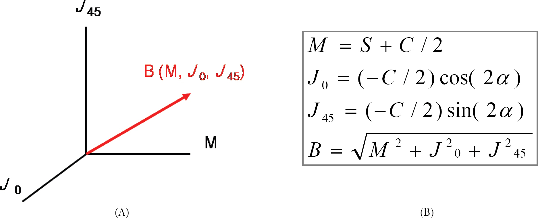
Figure 2. (A) The 3 Cartesian coordinates (x, y, z) of each power vector correspond to the powers of 3 lenses that, in combination, fulfill a refractive prescription: a spherical lens of power M, a Jackson crossed cylinder of power J0 with axes at 90 degrees and 180 degrees, and a Jackson crossed cylinder of power J45 with axes at 45 degrees and 135 degrees. The Pythagorean length of the power vector, B, is a measure of overall blurring strength of a spherocylindrical lens or refractive error. (B) Power vector analysis (S=spherical diopters, C=cylindrical diopters, α=axis (degree), Power vector=(M, J0, J45)).
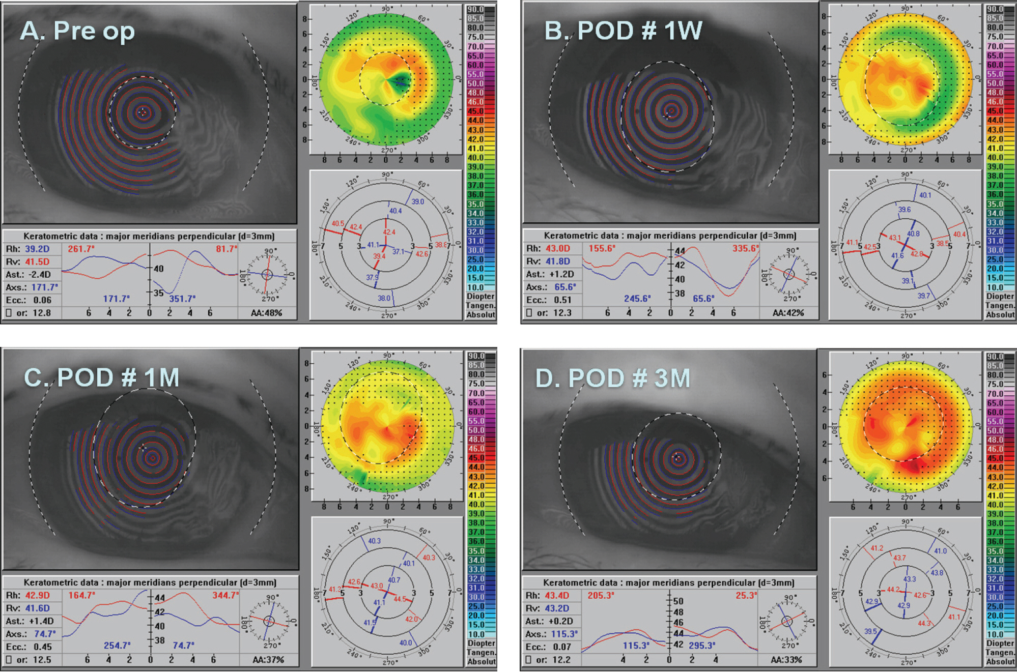
Figure 3. Pre- and post-operative topography pictures in a patient with pterygium. Astigmatism was with-the-rule preoperatively (A) but against-the-rule at the postoperative first week and postoperative third month (B)(C)(D).
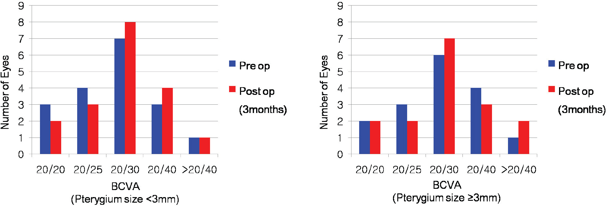
Figure 4. Pre- and post-operative best corrected visual acuity (BCVA) profile in patients. There was no significant change between pre- and postoperative BCVA in both <3 mm and ≥3 mm.
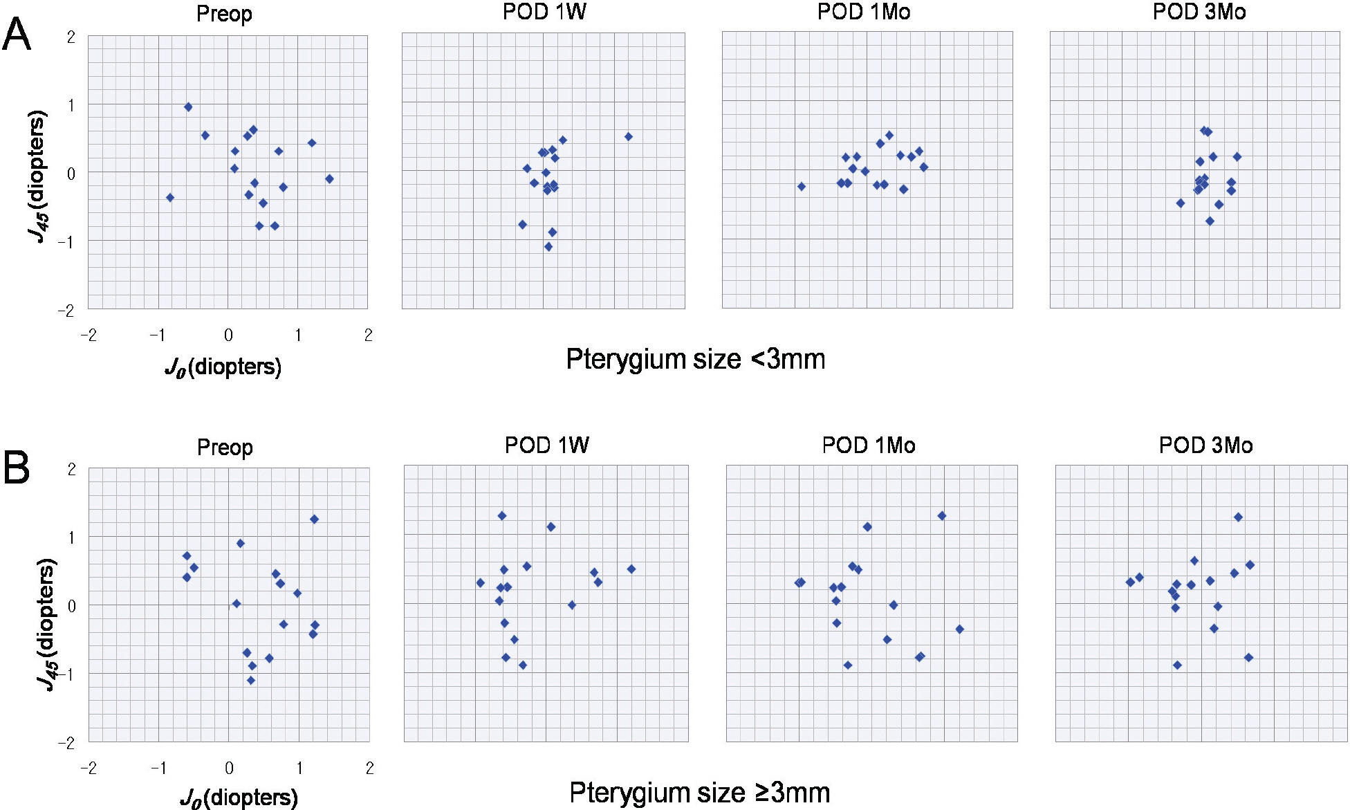
Figure 5. Scattergraph shows the astigmatic component of the power vector as represented by the 2-dimensional vector (J0, J45), which is the projection of the power vector into the astigmatism plane formed by the coordinate axes (J0, J45). Preoperative manifest astigmatism is reduced, and the cluster of points gathered around the zero point at postoperative third month for pterygium size <3 mm (A) but, the cluster of points did not gather for pterygium size ≥3 mm at postoperative third month (B).
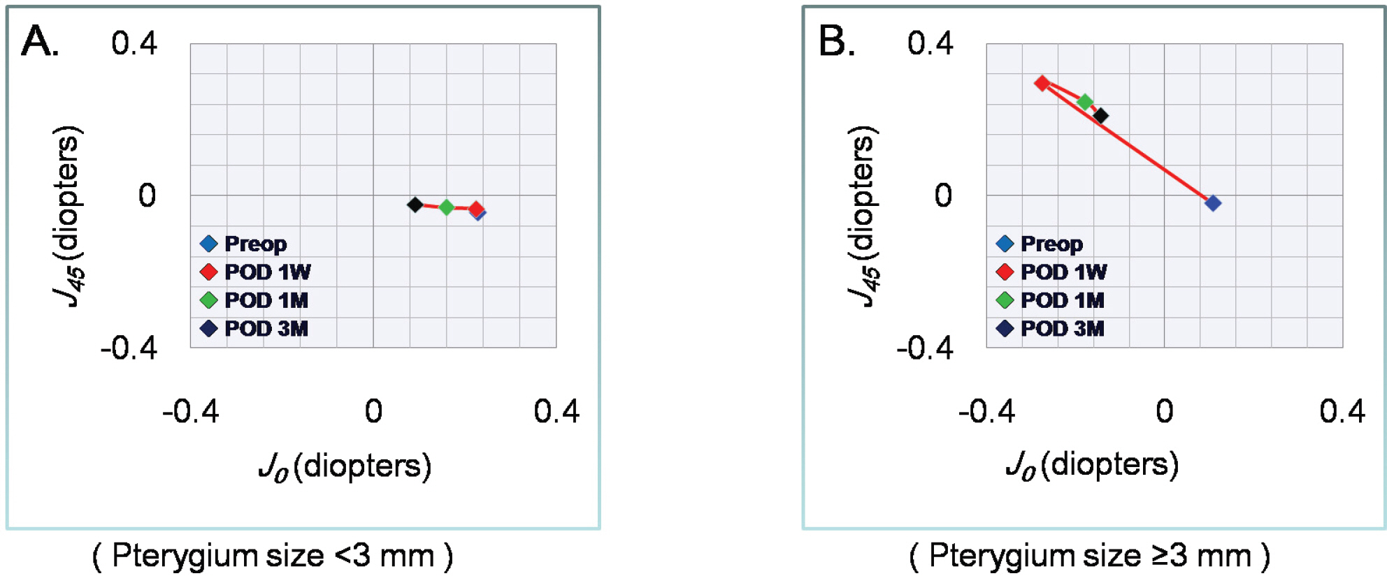
Figure 6. Scattergraph of power vector (J0, J45) on average shows that astigmatic axes did not change for pterygium size < 3 mm at the postoperative 3 months (A). But preoperative astigmatic axes changed from with-the-rule to against-the-rule at the postoperative first week for pterygium size ≥3 mm (B).
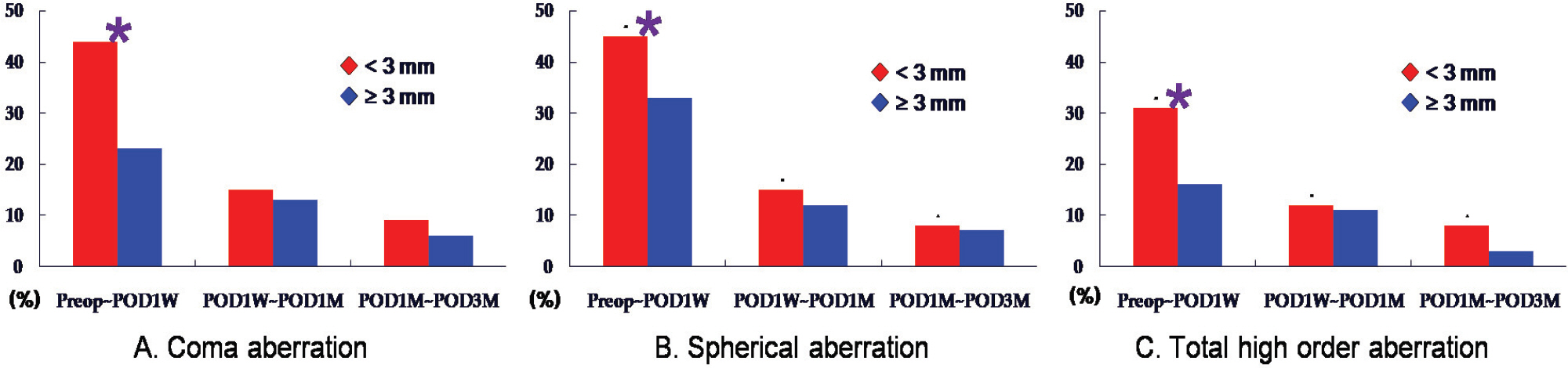
Figure 7. Change rates of high order aberrations (coma A, spherical B, total C) were significantly greater in group I than in group II at the preoperative period vs postoperative first week period (pterygium size <3 mm= group I, pterygium size ≥3 mm= group II), (* p<0.05, Wilcoxon signed rank test).
Reference
-
References
1. Reinstrow SD. The conjunctiva. Podos SM, Yanoff M, editors. Textbook of Ophthalmology, first ed. London: Mosby- Wolfe;1995. v. 1:p. chap. 2.2. Lindsay RG, Sullivan L. Pterygium-induced corneal astigma tism. Clin Exp Optom. 2001; 84:200–3.3. Avisar R, Loya N, Yassur Y, Weinberger D. Pterygium- induced corneal astigmatism. Isr Med Assoc J. 2000; 2:14–5.4. Kampitak K. The effect of pterygium on corneal astigmatism. J Med Assoc Thai. 2003; 86:16–23.5. Walland MJ, Stevens JD, Steele AD. The effect of recurrent pterygium on corneal topography. Cornea. 1994; 13:463–4.
Article6. Hansen A, Norn M. Astigmatism and surface phenomena in pterygium. Acta Ophthalmol. 1980; 58:174–81.
Article7. Gridley MJ, Perlman EM. A form of variable astigmatism induced by pseudopterygium. Ophthalmic Surg. 1986; 17:794–795.
Article8. Oldenburg JB, Garbus J, McDonnell JM, McDonnell PJ. Conjunctival pterygia: mechanism of corneal topographic changes. Cornea. 1990; 9:200–4.9. Kang SW, Cho BJ. Changes in the corneal curvature and recurrence rate following pterygium surgery with relation to pterygium size and morphology. J Korea Ophthalmol Soc. 2001; 42:1255–64.10. Soriano JM, Janknecht P, Witschel H. Effect of pterygium operation on preoperative astigmatism: prospective study. Ophthalmologe. 1993; 90:688–90.11. Budak K, Khater TT, Friedman NJ, Koch DD. Corneal topographic changes induced by excision of perilimbal lesions. Ophthalmic Surg Lasers. 1999; 30:458–64.
Article12. Stern GA, Lin A. Effect of pterygium excision on induced corneal topographic abnormalities. Cornea. 1998; 17:23–7.
Article13. Oh JR, Kim JS, Lee DH. The change of ocular aberration after LASIK surgery. J Korea Ophthalmol Soc. 2003; 44:278–83.14. Roberts C. The cornea is not a piece of plastic. J Refract Surg. 2000; 16:407–13.
Article15. Pesudovs K, Figueiredo FC. Corneal first surface wavefront aberrations before and after pterygium surgery. J Refract Surg. 2006; 22:921–5.
Article16. Oner FH, Kaderli B, Durak I, Cingil G. Analysis of the pterygium size inducing marked refractive astigmatism. Eur J Ophthalmol. 2000; 10:212–4.
Article17. Thibos LN, Horner D. Power vector analysis of the optical outcome of refractive surgery. J Cataract Refract Surg. 2001; 27:80–5.
Article18. Tomidokoro A, Miyata K, Sakaguchi Y, et al. Effects of pterygium on corneal spherical power and astigmatism. Ophthalmology. 2000; 107:1568–71.
Article19. Bahar I, Loya N, Weinberger D, Avisar R. Effect of pterygium surgery on corneal topography: a prospective study. Cornea. 2004; 23:113–7.20. Yagmur M, Ozcan AA, Sari S, Ersöz TR. Visual acuity and corneal topographic changes related with pterygium surgery. J Refract Surg. 2005; 21:166–70.
Article21. Lin A, Stern GA. Correlation between pterygium size and induced corneal astigmatism. Cornea. 1998; 17:28–30.
Article22. Ozdemir M, Cinal A. Early and late effects of pterygium surgery on corneal topography. Ophthalmic Surg Laser Imaging. 2005; 36:451–6.
Article23. Hong JW, Lee TS. Comparison of refractive change measured by corneal topography between before and after pterygium excision. J Korea Ophthalmol Soc. 1996; 37:1614–9.24. Maheshwari S. Pterygium-induced corneal refractive changes. Indian J Ophthalmol. 2007; 55:383–6.
Article25. Avisar R, Loya N, Yassur Y, Weinberger D. Pterygium- induced corneal astigmatism. Isr Med Assoc J. 2000; 2:14–5.26. Smolek MK, Klyce SD. Zernike polynomial fitting fails to represent all visually significant corneal aberrations. Invest Ophthalmol Vis Sci. 2003; 44:4676–81.
Article27. Klyce SD, Karon MD, Smolek MK. Advantages and disadvantages of the Zernike expansion for representing wave aberration of the normal and aberrated eye. J Refract Surg. 2004; 20:S537–41.
Article
- Full Text Links
-
- Actions
-
Cited
- CITED
-
- Close
- Share
-
- Similar articles
-
- The Changes of Corneal Higher-Order Aberrations after Surgery According to Pterygium Size
- Usefulness of Refractive Measurement by Wavefront Aberrometer in Children
- Accuracy of the Hand-held Wavefront Aberrometer in Measurement of Refractive Error
- Comparison of Corneal Astigmatism and Higher-order Aberrations between Color Light-emitting Diode Topographer and Scheimpflug Imager
- The Change of Corneal Refractive Power Measured by Corneal Topography before and after Pterygium Surgery
