Changes of Anterior Chamber Depth and Angle After Cataract Surgery Measured by Anterior Segment OCT
- Affiliations
-
- 1Department of Ophthalmology, KyungHee University College of Medicine, Seoul, Korea. khjinmd@khmc.or.kr
- 2Department of Ophthalmology, Kangwon University College of Medicine, Gangwon, Korea.
Abstract
- PURPOSE
To report the change of anterior chamber parameters according to cataract severity after cataract surgery and to determine its relationship to the severity of cataract by using anterior segment optical coherence tomography.
METHODS
We measured the anterior chamber parameters in 19 eyes of 14 patients before, 1 week after, and 1 month after cataract surgery by slit lamp-adapted optical coherence tomography (SL-OCT). The measured parameters were as follows : the anterior chamber depth (ACD), the angle-opening distance 250 micrometer from the scleral spur (AOD250), the angle-opening distance 500 micrometer from the scleral spur (AOD500), and the trabecular-iris angle (TIA). We analyzed the relationship between the severity of cataract and the change of the anterior chamber parameters.
RESULTS
The ACD, AOD250, AOD500, and TIA increased significantly at postoperative 1 week (P=0.000, 0.002, 0.005, 0.022) and 1 month (P=0.000, 0.004, 0.001, 0.002). The preoperative parameters were negatively correlated with the differences between the postoperative 1 week and preoperative parameters (gamma=-0.834, -0.591, -0.421, -0.826) and between postoperative 1 month and preoperative parameters (gamma=-0.659, -0.700, -0.770, -0.821). The change of parameters at postoperative 1 week (by N P=0.959, 0.916, 0.824, 1.000, by C P=0.454, 0.665, 0.578, 0.578) and 1 month (by N P=0.858, 0.973, 0.959, 0.959, by C P=0.999, 0.207, 0.950, 0.981) were not significantly different according to the severity of cataract (N, C).
CONCLUSIONS
Our results showed that cataract surgery significantly deepened the anterior chamber and widened its angle. The shallower and narrower the preoperative anterior chamber depth and angle were, respectively, the greater the postoperative changes of anterior chamber depth and angle were.
Keyword
Figure
-
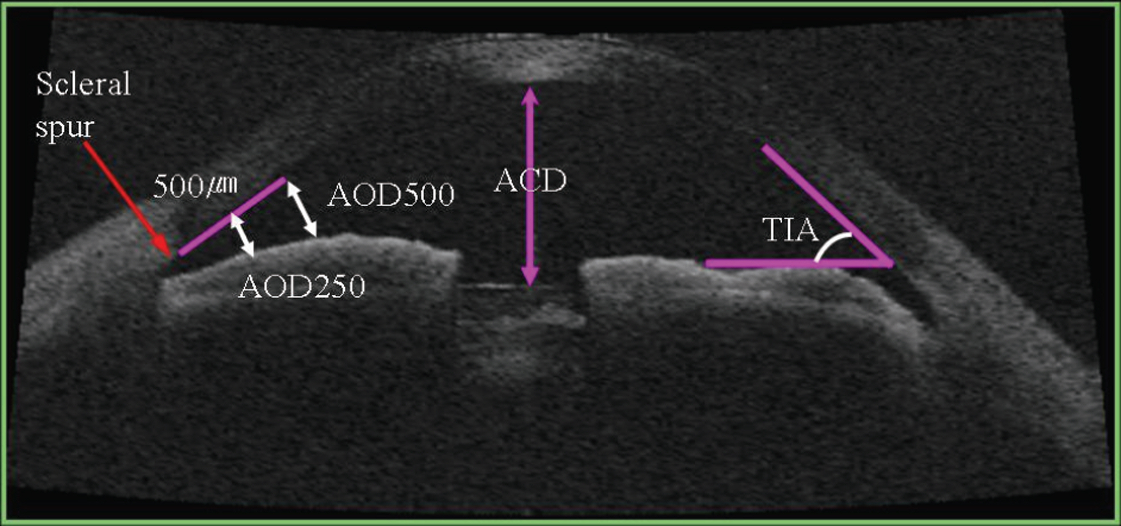
Figure 1. The definition of anterior chamber parameters. The anterior chamber depth (ACD) can be measured between the endothelium and the lens surface. The angle-opening distance was measured on a line perpendicular to the trabecular meshwork at points 250 µm (AOD250) and 500 µm (AOD500) from the scleral spur. The trabecular-iris angle was measured with the apex in the iris recess and the arms of the angle passing through a point on the trabecular meshwork 500 µm from the scleral spur and the point on the iris perpendicularly opposite.

Figure 2. The anterior segment OCT (optical coherence tomography) image by slit-lamp-adapted OCT and anterior chamber parameters of the anterior chamber in phakic eye (A) and pseudophakic eye (B). ACD1, which is measured between the inner corneal surface and the intraocular lens surface, extends into the posterior chamber (Pavlin’s original definition). ACD2, which is measured between the inner corneal surface and the plane of the posterior aspect of the iris at pupillary margin, represents the real anterior chamber depth in eyes with pseudophakia. As ACD1 extends into the posterior chamber, it is not the real ACD in pseudophakia. AOD250, which is the angle-opening distance at 250 µm from the scleral spur. AOD500, which is the angle-opening distance at 500 µm from the scleral spur. TIA, which is the trabecular-iris angle.

Figure 3. Anteior segment optical coherence tomography (OCT) image before cataract surgery (A), postoperative 1 week (B), postoperative 1 month (C). Note deepening of the anterior chamber depth (ACD), flattening of the convex iris configuration, and widening of the angle after cataract surgery.
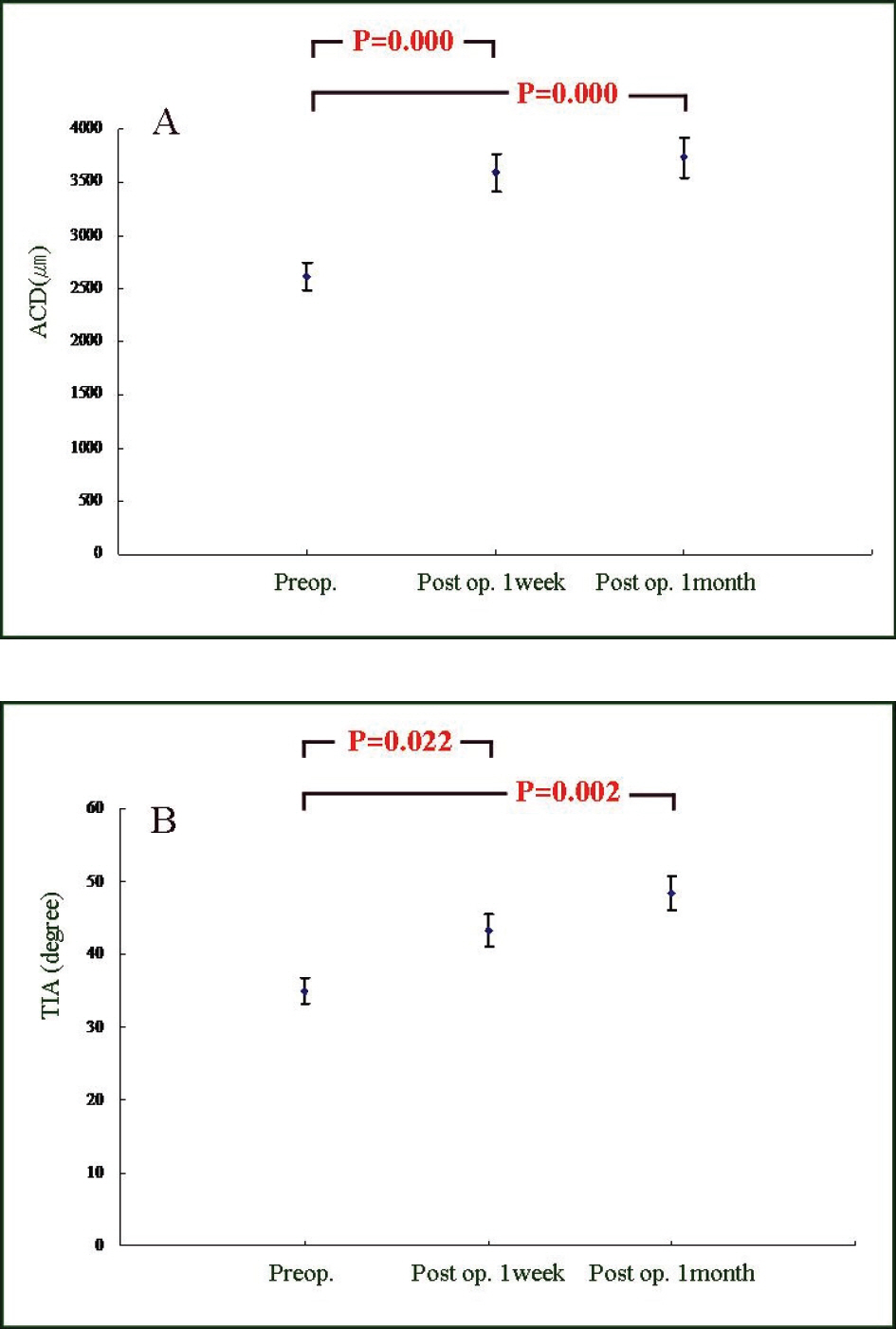
Figure 4. The changes of anterior chamber parameters before and after surgery. (A) ACD change. (B) TIA change. (ACD, anterior chamber depth; TIA, trabecular-iris angle; P, p-value by Wilcoxon sign rank test)
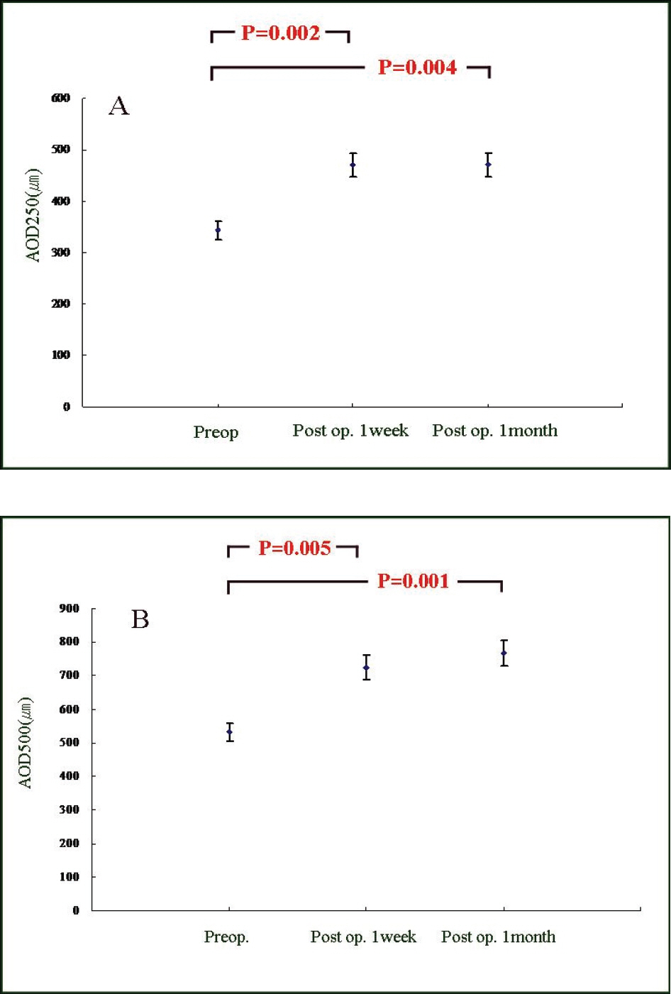
Figure 5. The changes of anterior chamber parameters before and after surgery. (A) AOD250 change. (B) AOD500 change. (AOD250, angle-opening distance at 250 µm from scleral spur; AOD500, angle-opening distance at 500 µm from scleral spur; P, p-value by Wilcoxon sign rank test)

Figure 6. The preoperative anterior chamber parameters and ratio of postoperative 1 week parameters. X-axis: preoperative value, Y-axis: postoperative 1 week value/preoperative value (A) ACD (B) AOD250 (C) AOD500 (D) TIA. (ACD, anterior chamber depth; AOD250, angle-opening distance at 250 µm from scleral spur; AOD500, angle-opening distance at 500µm from scleral spur, TIA, trabecular-iris angle)
Reference
-
References
1. Steuhl KP, Marahrens P, Frohn C, Frohn A. Intraocular pressure and anterior chamber depth before and after extracapsular cataract extraction with posterior chamber lens implantation. Ophthalmic Surg. 1992; 23:233–7.
Article2. Koo BS, Chung J, Baek NH. The effect of extracapsular cataract extraction in patients with chronic angle-closure glaucoma combined with cataract. J Korean Ophthalmol Soc. 1996; 37:1045–53.3. Kurimoto Y, Park M, Sakaue H, Kondo T. Changes in the anterior chamber configuration after small-incision cataract surgery with posterior chamber intraocular lens implantation. Am J Ophthalmol. 1997; 124:775–80.
Article4. Lee HJ, Chung SK, Baek NH. Changes of preoperative and postoperative anterior chamber angle in phacoemulsification and planned extracapsular cataract extraction. J Korean Ophthalmol Soc. 1998; 39:1170–5.5. Pereira FA, Cronemberger S. Ultrasound biomicroscopic study of anterior segment changes after phacoemulsification and foldable intraocular lens implantation. Ophthalmology. 2003; 110:1799–806.
Article6. Nonaka A, Kondo T, Kikuchi M. . Angle widening and alteration of ciliary process configuration after cataract surgery for primary angle closure. Ophthalmology. 2006; 113:437–41.
Article7. Memarzadeh F, Tang M, Li Y. . Optical coherence tomography assessment of angle anatomy changes after cataract surgery. Am J Ophthalmol. 2007; 144:464–5.
Article8. Shibata T, Hockwin O, Weigelin E. . Biometry of the lens with respect to age and cataract morphology. Evaluation of Scheimpflug photos of the anterior segment. Klin Monatsbl Augenheilkd. 1984; 185:35–42.9. Richard DW, Russell SR, Anderson DR. A method for improved biometry of the anterior chamber with a Scheimpflug technique. Invest Ophthalmol Vis Sci. 1988; 29:1826–35.10. Shibata T, Sazuki K, Skamoto Y, Takahashi N. Quantitative chamber angle measurement utilizing image-processing techniques. Ophthalmic Res. 1990; 22:–S81. 4
Article11. Pavlin CJ, Harasiewiez K, Foster FS. Ultrasound biomicroscopy of anterior segment structures in normal and glaucomatous eyes. Am J Ophthalmol. 1992; 113:381–9.
Article12. Wirbelauer C, Gochmann R, Pham DT. Imaging of the anterior eye chamber with optical coherence tomography. Klin Monatsbl Augenheilkd. 2005; 222:856–62.13. Radhakrishnan S, Goldsmith J, Huang D. . Comparison of optical coherence tomography and ultrasound biomicroscopy for detection of narrow anterior chamber angles. Arch Ophthalmol. 2005; 123:1053–9.
Article14. Müler M, Dahmen G, Pörksen E. . Anterior chamber angle measurement with optical coherence tomography: Intraobserver and interobserver variability. J Cataract Refract Surg. 2006; 32:1803–8.15. Murphy GE. Long-term gonioscopy follow-up of eyes with posterior lens implants and no iridectomy. Ophthalmic Surg. 1986; 17:227–8.16. Huang D, Swanson EA, Lin CP. . Optical coherence tomography. Science. 1991; 254:1178–81.
Article17. Radhakrishnan S, Huang D, Smith SD. Optical coherence tomography imaging of anterior chamber angle. Ophthalmol Clin North Am. 2005; 18:375–81.18. Lee JH, Park WC, Rho SH. The effects of pilocarpine on the anterior chamber depth and angle. J Korean Ophthalmic Soc. 1994; 35:572–9.19. Arai M, Ohzuno I, Zako M. Anterior chamber depth after posterior chamber intraocular lens implantation. Acta Ophthalmol. 1994; 72:694–7.
Article20. Yoshida S, Hashiba H, Tsukuda M, Ohara Y. Significance of angle of intraocular lens haptics on anterior chamber depth. Jpn J Clin Ophthalmol. 1989; 43:173–6.
- Full Text Links
-
- Actions
-
Cited
- CITED
-
- Close
- Share
-
- Similar articles
-
- A Study for Measurement of the Anterior Chamber Depth and Angle Using Image Analysis Technique in Cataractous Eyes
- Measurement of Anterior Segment Using Visante OCT in Koreans
- Changes in Anterior Chamber Depth and Angle After Phacoemulsification measured by Anterior Segment Optical Coherence Tomography
- The Effect of Extracapsular Cataract Extraction in Patients with Chronic Angle-Closure Glaucoma combined with Cataract
- Diurnal Variation in the Depth of the Anterior Chamber and Correlation between the Depth of the Anterior Chamber and Intraocular Pressure
