J Korean Ophthalmol Soc.
2008 Sep;49(9):1431-1436.
Clinical Manifestations of Avellino Corneal Dystrophy Diagnosed by Non-invasive Genetic Test
- Affiliations
-
- 1Department of Ophthalmology, Korea University College of Medicine, Seoul, Korea. crisim@korea.ac.kr
Abstract
- PURPOSE
To introduce a new genetic method for the diagnosis of Avellino corneal dystrophy (ACD), which is non-invasive and can be easily performed on an outpatient basis, and to evaluate the relationship between the degree of corneal opacity and age or sex.
METHODS
A genetic study was performed on 11 patients who had a specific corneal opacity by slit-lamp examination and on four normal patients by using a specific adhesive tape to obtain epidermal keratinocytes. Corneal dystrophy was diagnosed according to the genetic study.
RESULTS
All 11 patients were confirmed as having heterozygous ACD. Heterozygous ACD patients were classified into five stages: trace, mild, moderate, severe, or very severe, based on slit-lamp photography status. Corneal stages had no relationship with sex (p=0.982), but the severity of ACD increased with age (p=0.005).
CONCLUSIONS
A non-invasive sticker-type genetic study kit, the "U-gene test" is a good method to diagnose corneal dystrophy genetically. Avellino corneal dystrophy becomes more severe over time but has no relationship with sex.
MeSH Terms
Figure
-

Figure 1. Showing how to use “U-gene test.” Put the sticker on the forearm about 20 seconds and peel it off.

Figure 2. Index photographs of Avellino corneal dystrophy. Five stages based on a slit-lamp photography. (A) Trace, (B) Mild, (C) Moderate, (D) Severe, (E) Very Severe.
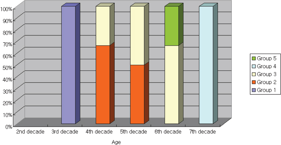
Figure 3. Distribution of heterozygous Avellino corneal dystrophy according to the age. ACD aggravates as the patients become older.
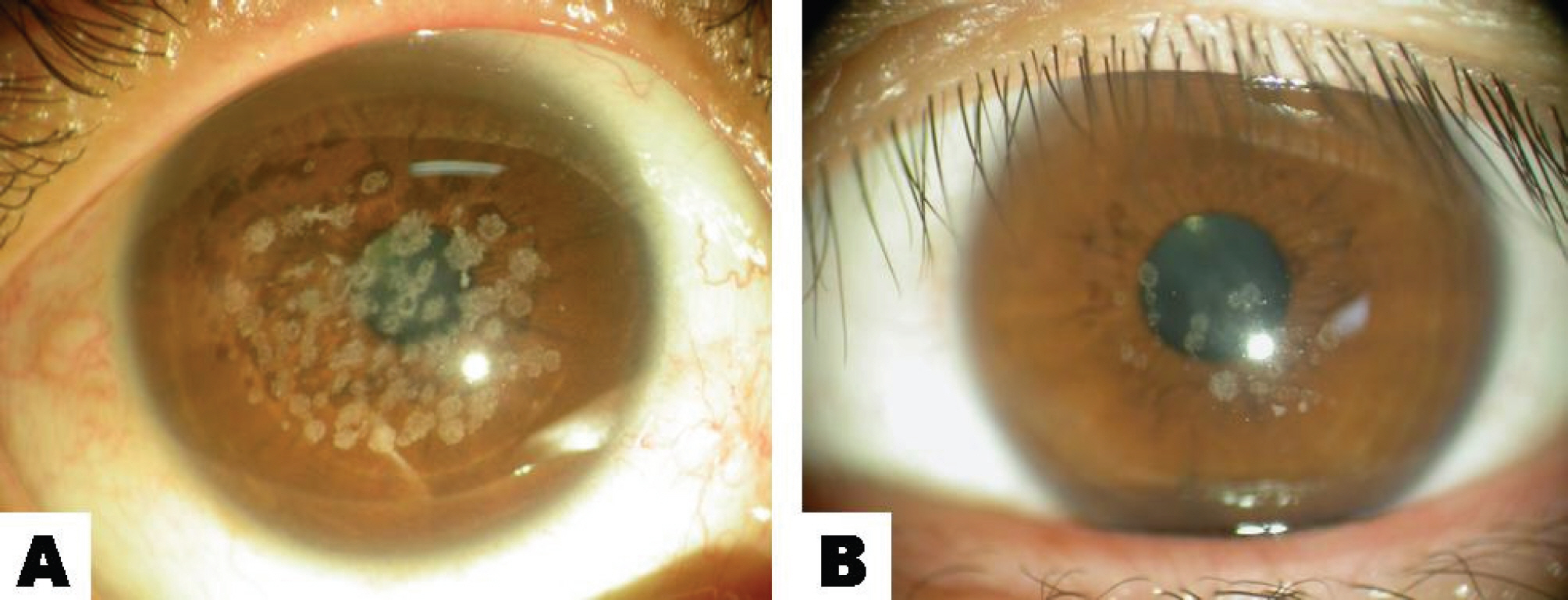
Figure 5. A slit lamp photograph showing the different stages of Avellino corneal dystrophy in the patients of similar age. (A) A 48-year-old female (B) A 42-year-old female.
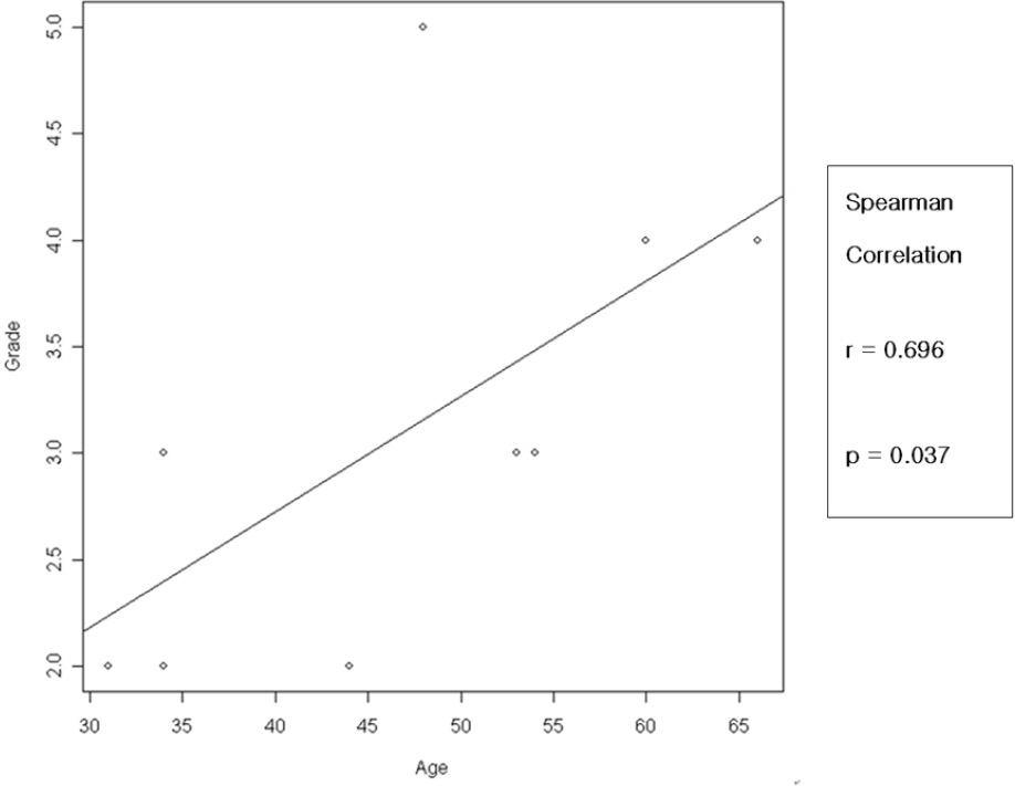
Figure 6. Linear correlation between ACD grade and age after excluding one of the family members.
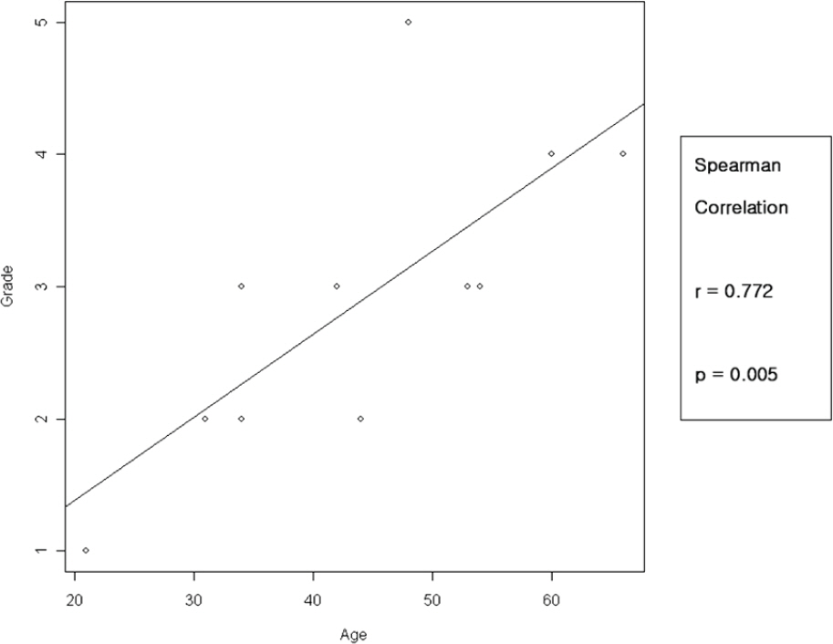
Figure 4. Linear correlation between ACD grade and age.
Reference
-
References
1. Lisch W. Classification of corneal dystrophies. Ophthalmic Res. 2006; 38:176.
Article2. Pieramici SF, Afshari NA. Genetics of corneal dystrophies: the evolving landscape. Curr Opin Ophthalmol. 2006; 17:361–6.
Article3. Ye YF, Yao YF, Zhou P, Pan F. In vivo confocal microscopy of pre-Descemet's membrane corneal dystrophy. Clin Experiment Ophthalmol. 2006; 34:614–6.
Article4. Konishi M, Mashima Y, Nakamura Y. . Granular-lattice (Avellino) corneal dystrophy in Japanese patients. Cornea. 1997; 16:635–8.
Article5. Kim HS, Yoon SK, Cho BJ. . BIGH3 gene mutations and rapid detection in Korean patients with corneal dystrophy. Cornea. 2001; 20:844–9.
Article6. Kocak-Atlintas AG, Kocak-Midillioglu I, Akarsu AN, Duman S. BIGH3 gene analysis in the different diagnosis of corneal dystrophies. Cornea. 2001; 20:64–8.7. Klintworth GK. Advances in the molecular genetics of corneal dystrophies. Am J Ophthalmol. 1999; 128:747–54.
Article8. Waring GO, Rodringues MM, Labison PR. Corneal dystrophies. Surv Ophthalmol. 1978; 23:71–122.9. Cogan DG, Donaldson DD, Kuwabara T, Marshall D. Microcystic dystrophy of the corneal epithelium. Trans Am Ophthalmol Soc. 1964; 63:213–25.10. Ferry AP, Benson WH, Weinberg RS. Combined granular- lattice (Avellino) corneal dystrophy. Trans Am Ophthalmol Soc. 1997; 95:61–77.11. Jee DH, Lee YD, Kim MS. Epidemiology of corneal dystrophy in Korea. J Korean Ophthalmol Soc. 2003; 44:581–7.12. Chung SH, Kim CY, Kim EK. The classification and clinical characteristics in Korean Patients with Avellino Corneal Dystrophy. J Korean Ophthalmol Soc. 2005; 46:938–44.13. Roh MI, Grossniklaus HE, Chung SH. . Avellino corneal dystrophy exacerbated after LASIK: scanning electron microscopic findings. Cornea. 2006; 25:306–11.14. Jun RM, Tchah H, Kim TI. . Avellino corneal dystrophy after LASIK. Ophthalmology. 2004; 111:463–8.
Article
- Full Text Links
-
- Actions
-
Cited
- CITED
-
- Close
- Share
-
- Similar articles
-
- Phototherapeutic Keratectomy of Avellino Corneal Dystrophy with 0.02% Mitomycin C
- Confocal Microscopic Findings of Avellino Corneal Dystrophy According to Disease Severity
- Four Cases of Avellino Corneal Dystrophy Concurrent with Floppy Eyelid Syndrome
- The Classification and Clinical Characteristics in Korean Patients with Avellino Corneal Dystrophy
- A Case of Spontaneous Regression of Schnyder's Crystalline Corneal Dystrophy
