J Korean Ophthalmol Soc.
2007 Sep;48(9):1285-1290.
A Case of Hallermann-Streiff Syndrome
- Affiliations
-
- 1Department of Ophthalmology, Maryknoll Hospital, Pusan, Korea. wansookim@yahoo.com
Abstract
-
PURPOSE: We report a case of Hallermann-Streiff syndrome with 360 degrees posterior synechiae, small pupils and aphakia.
METHODS
A five-year-old female presented with decreasing visual acuity of both eyes. Visual acuity was not checkable due to mental retardation. Microcornea, microphthalmia, nystagmus and esotropia were found, and a fundus examination was not available due to 360 degrees posterior synechiae and small pupils. She had developmental delays, bird-like face and hypotrichosis. A pediatric physician was consulted who diagnosed her with Hallermann-Streiff syndrome. Refraction and fundus examinations were impossible due to her small pupils, so synechiolysis was done.
RESULTS
After synechiolysis and pupil dilatation in right eye with iris retractors, continuous curvilinear capsulorhexis (CCC) was attempted. However, the anterior capsule was unusually fragile and fibrtic. Therefore, the CCC failed. In addition, the crystalline lens and the zonule were not found. The posterior capsule was fragile similar to the anterior capsule. Complete posterior CCC (PCCC) was impossible. We could not find any formed vitreous in the vitreous cavity during anterior vitrectomy. We diagnosed the condition as aphakia with only two layers of membranes. Two weeks later, synechiolysis in the left eye was done. The left eye was also diagnosed with aphakia, and only synechiolysis was performed.
CONCLUSIONS
The possibility of aphakia must be always considered in cases of Hallermann-Streiff syndrome.
MeSH Terms
Figure
-

Figure 1. Preoperative findings show a small pupil with 360-degree posterior synechiae. (A) Right eye. (B) Left eye.
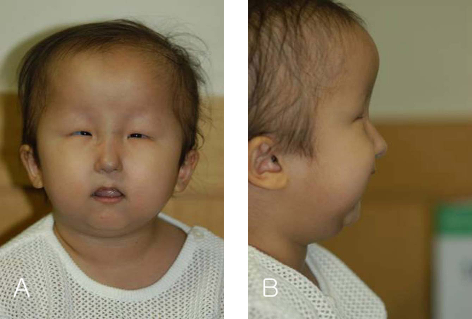
Figure 2. (A) Frontal view of the face shows a bird-like face with a long nose and microophthalmia. (B) Side view of the face shows frontal bossing and emphasizes the bird-like face with underdevelopment of the mandible.
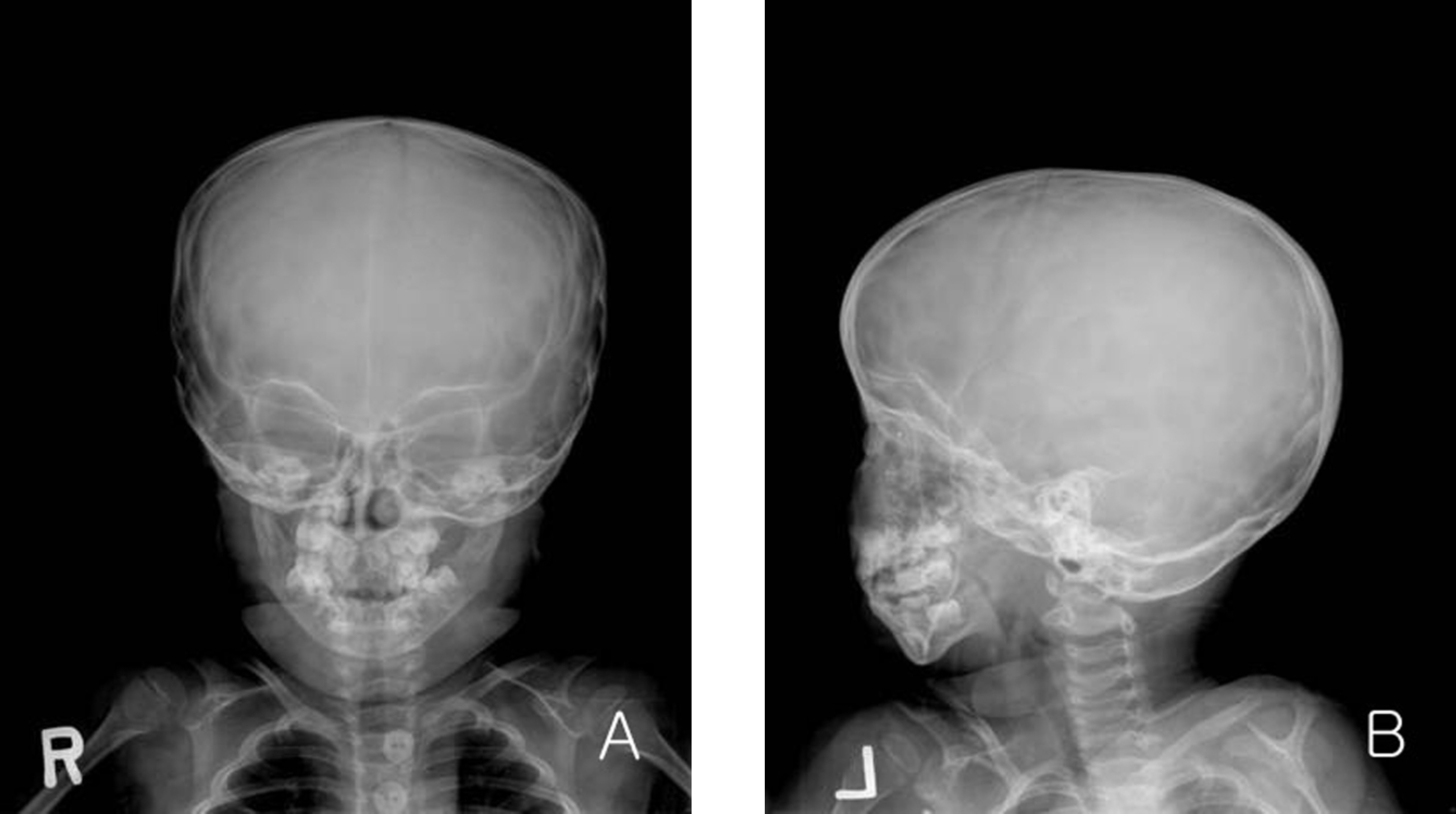
Figure 3. X-ray findings of the skull show a hypoplastic mandible and small orbits. Cranio-facial ratio increased twofold. (A) The antero-posterior view. (B) The left lateral view.
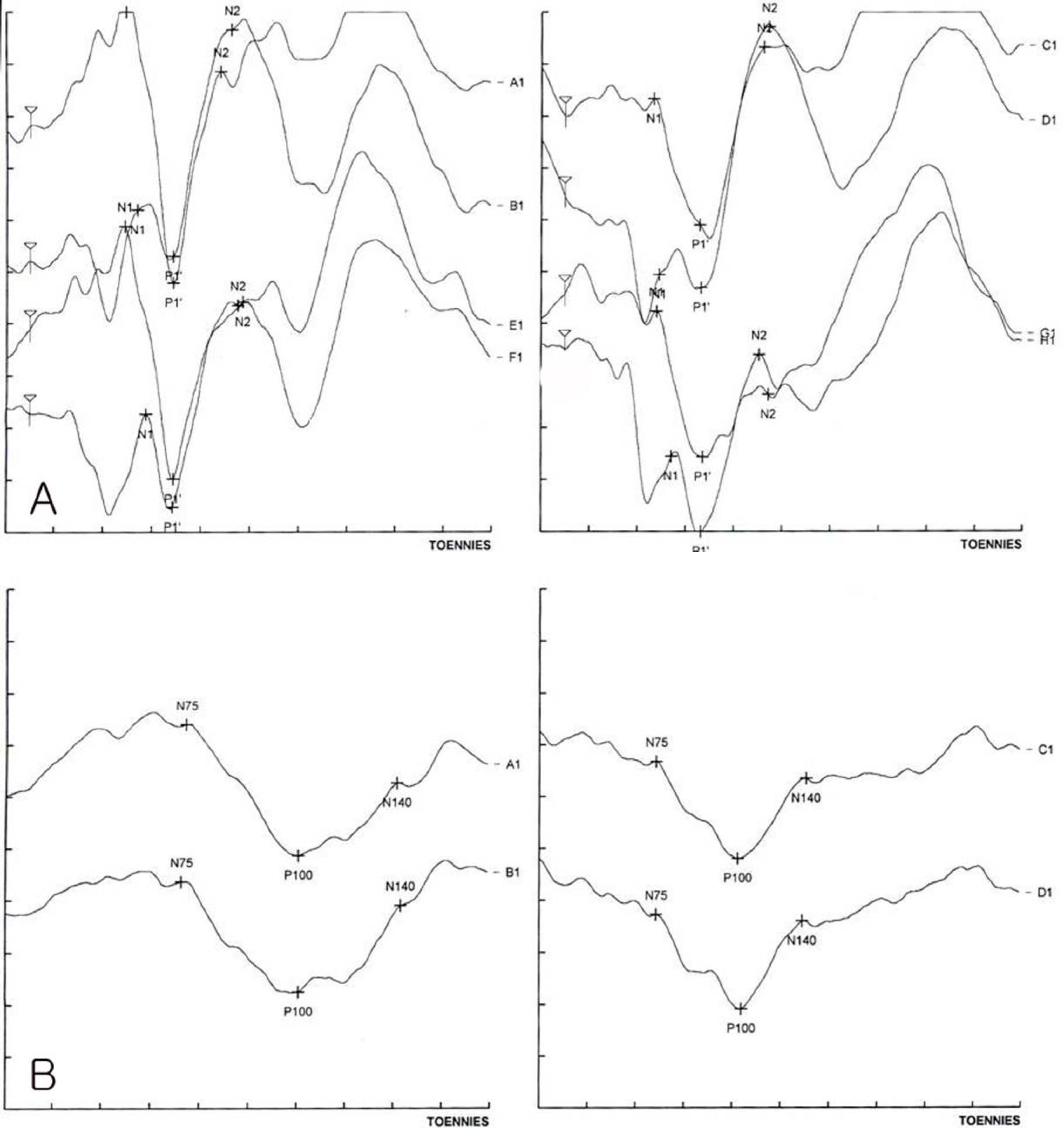
Figure 4. (A) Latencies of P1 wave were prolonged in right and left flash visual evoked potential (VEP). (B) Latencies of P100 value were prolonged in right and left pattern VEP.
Reference
-
References
1. Hallermann W. Vogelgescht und Cataracta Congenita. Klin Monatsbl Augenh. 1948; 113:315–8.2. Striff EB. Dysmorphie Mandibulfaciale (tefed’ Oiseall) et alterations oculaire. Ophthalmologica. 1950; 120:79–83.3. Son JS, Hwang HY, Moon HK, Hah JO. A Case of Hallermann-Streiff Syndrome. J Korean Pediatr Soc. 1987; 30:691–4.4. Kim SY, Kim YM, Yoon HS. A Case of Hallermann-Streiff Syndrome with Intra-Uterine Growth Retardation. J Korean Pediatr Soc. 2003; 46:926–9.5. Koo BS, Rhee SU. A Case of Hallermann-Streiff Syndrome. J Korean Ophthalmol Soc. 1967; 8:55–60.
Article6. Shim WS, Kim KR. A Case of Hallermann-Streiff Syndrome. J Korean Ophthalmol Soc. 1976; 17:297–9.
Article7. Lee HB, Kim JH. A Case of Hallermann Streiff Syndrome. J Korean Ophthalmol Soc. 1978; 19:455–8.8. Ryoo MH, Kim SS, Yi KP. A Case of Hallermann-Streiff Syndrome. J Korean Ophthalmol Soc. 1990; 31:831–6.
Article9. Francois J. A New Syndrome: dyscephalia with bird face and dental anomalies, nanism, hypotrichosis, cutaneous atrophy, microphthalmia and congenital cataract. AMA Arch Ophthalmol. 1958; 60:842–62.10. Schanzlin DJ, Goldberg DB, Brown SI. Hallermann-Streiff Syndrome associated with sclerocornea, aniridia, and a chromosomal abnormality. Am J Ophthalmol. 1980; 90:411–5.
Article11. Hoefnagel D, Benirchke K. Dyscephalia mandibulooculofacialis (Hallermann-Streiff syndrome). Arch Dis Child. 1965; 40:57–61.
Article12. Ide CH, Webb RW. Hallerman-Streiff syndrome. Am J Ophthalmol. 1969; 67:151–3.13. Steele RW, Bass JW. Hallermann-Streiff syndrome. Am J Dis Child. 1970; 120:462–5.
Article14. Golomb RS, Porter PS. A distinct hair shaft abnormality in the Hallermann-Streiff syndrome. Cutis. 1975; 16:122–8.15. Kurlander GJ, Lavy NW, Campbell JA. Roentgen differentiation of the oculudentodigital syndrome and the Hallermann-Streiff syndrome in infancy. Radiology. 1969; 86:77–85.16. Caspersen I, Warberg M. Hallermann-Streiff Syndrome. Acta Ophthalmol. 1968; 46:385–90.
Article17. Wright KW, Spiegel PH. Pediatric Ophthalmology and Strabismus. 2nd ed.New York: Springer;2002. p. 725–7.18. Hopkins DJ, Horan EC. Glaucoma in the Hallermann-Streiff Syndrome. Br J Ophthalmol. 1970; 416–22.
Article19. Cho KS, Shin YB, Kim BC. Interrelationship between Axial Length and Refractive States, and Anterior Chamber Depth in the Newborn. J Korean Ophthalmol Soc. 1990; 31:215–9.20. Kim JH, Lee SH. Intraocular Pressure, Corneal Diameter and C/D Ratio in Normal Newborns. J Korean Ophthalmol Soc. 1996; 37:115–8.21. Wolter JR, Jones DH. Spontaneous Cataract Absorption in Hallermann-Streiff Syndrome. Ophthalmologica. 1965; 150:401–8.
Article22. Sato M, Amano E, Okamoto Y, Miyake Y. Ultrasound Bimicroscopic Findings in Hallermann-Streiff Syndrome. Jpn J Ophthalmol. 2002; 46:451–4.23. Aracena T, Sanguesa P. Hallermann-Streiff-Francois syndrome. J Pediatr Ophthalmol. 1977; 14:373–8.
Article24. Belkin M, Ticho U, Susal A, Levinson A. Ultrasonography in the refraction of aphakic infants. Br J Ophthalmol. 1973; 57:845–8.
Article
