J Korean Rheum Assoc.
2010 Sep;17(3):331-332.
A Case of Systemic Amyloidosis
- Affiliations
-
- 1Department of Internal Medicine, Dongsan Medical Center, Keimyung University School of Medicine, Daegu, Korea. mdkim9111@hanmail.net
Abstract
- No abstract available.
MeSH Terms
Figure
-
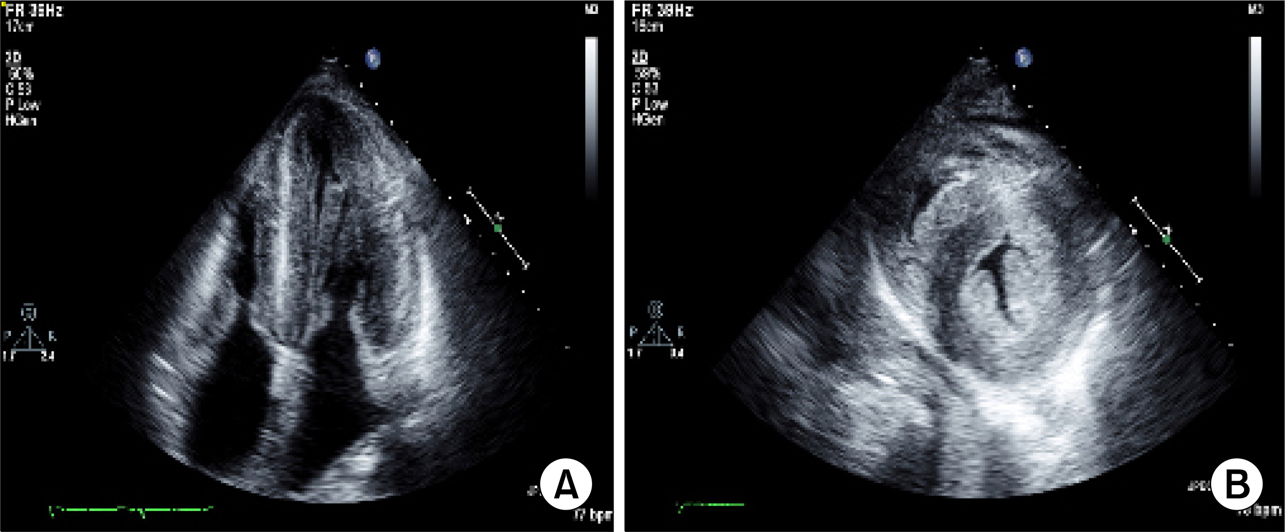
Fig. 1. (A, B) Concentric hypertrophy, thickened septum and ventricular wall with characteristic granular sparkling and pericardial effusion.

Fig. 2. Polarizing illumination after Congored staining showed typical green birefringence amyloid fibril (Congored) sural nerve biopsy (A), abdominal fat biopsy (B).
Reference
-
1). Glenner GG. Amyloid deposits and amyloidosis. The beta-fibrilloses (first of two parts). N Engl J Med. 1980. 302:1283–92.2). Lee SW., Lee JH., Kim KH., Chung WT. Clinical manifestations of secondary amyloidosis associated with rheumatoid arthritis. J Korean Rheum Assoc. 2004. 11:326–32.3). Kim TJ., Oh SI., Park JS., Kim TY., Park JH., Uhm WS, et al. The secondary amyloidosis in rheumatic diseases. J Korean Rheum Assoc. 2003. 10:39–44.
