Effect of perioperative buccal fracture of the proximal segment on postoperative stability after sagittal split ramus osteotomy
- Affiliations
-
- 1Department of Oral and Maxillofacial Surgery, Seoul National Unversity Dental Hospital, School of Dentistry, Brain Korea Plus, Seoul National Unversity, Seoul, Korea. sjhwang@snu.ac.kr
- 2Dental Research Institute, Seoul National University, Seoul, Korea.
Abstract
OBJECTIVES
Buccal fracture of the mandibular proximal bone segment during bilateral sagittal split ramus osteotomy (SSRO) reduces the postoperative stability. The primary aim of this study is to evaluate the effect of this type of fracture on bone healing and postoperative stability after mandibular setback surgery.
MATERIALS AND METHODS
Ten patients who experienced buccal fracture during SSRO for mandibular setback movement were evaluated. We measured the amount of bone generation on a computed tomography scan, using an image analysis program, and compared the buccal fracture side to the opposite side in each patient. To investigate the effect on postoperative stability, we measured the postoperative relapse in lateral cephalograms, immediately following and six months after the surgery. The control group consisted of ten randomly-selected patients having a similar amount of set-back without buccal fracture.
RESULTS
Less bone generation was observed on the buccal fracture side compared with the opposite side (P<0.05). However, there was no significant difference in anterior-posterior postoperative relapse between the group with buccal fracture and the control group. The increased mandibular plane angle and anterior facial height after the surgery in the group with buccal fracture manifested as a postoperative clockwise rotation of the mandible.
CONCLUSION
Bone generation was delayed compared to the opposite side. However, postoperative stability in the anterior-posterior direction could be maintained with rigid fixation.
Keyword
Figure
-
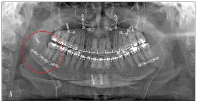
Fig. 1 Rigid fixation of a buccal fracture of the proximal segment. The buccal fracture on the right side was stabilized using long mini-plate and monocortical screws and additional bicortical positional screw (circle).
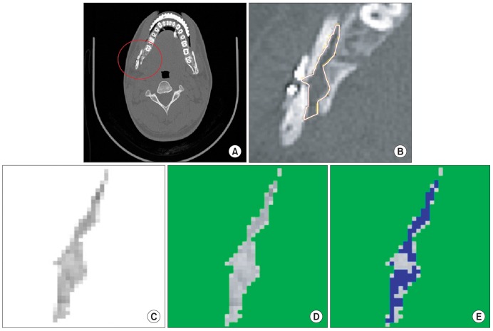
Fig. 2 Evaluation of bone regeneration using Image J. A. The buccal fracture was occurred on right proximal segment (circle). B. The total inter-segmental area between the proximal and distal segments was defined based on the axial computed tomography image. C. The total inter-segmental area was adjusted to the threshold below 210 in the Image J program, where the threshold of cortical and medullar bone was greater than 210. D, E. The area of newly formed bone (threshold from 190 to 210 in the Image J program) was extracted from the total inter-segmental area. Soft tissue and dead space (threshold below 190) were also removed. Finally, the ratio of new bone area to total inter-segmental area was calculated.
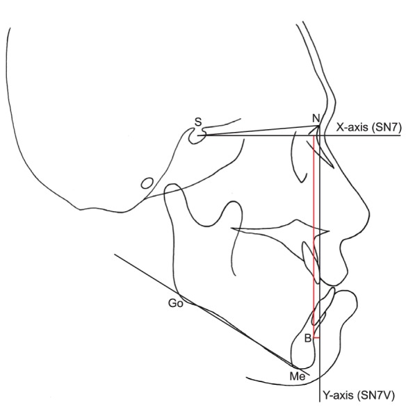
Fig. 3 Post-operative stability was evaluated by measuring the changes in reference points and reference planes: sella (S), nasion (N), x-axis (SN7, a line drawn 7° lower to the S-N line), y-axis (SN7V, a line on the N and perpendicular to SN7), point B (B), menton (Me), gonion (Go).
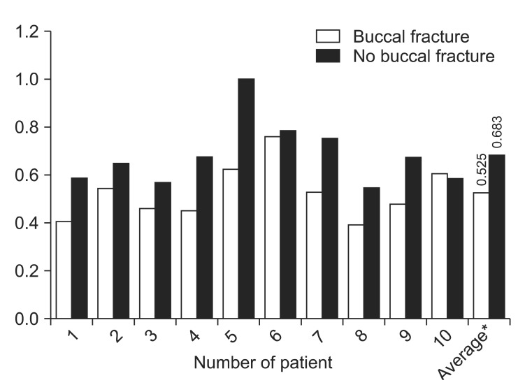
Fig. 4 Comparison of new bone formation between the buccal fracture side and non-fractured side. Bone healing was significantly delayed on the fractured side compared with the opposite side. *P=0.019.
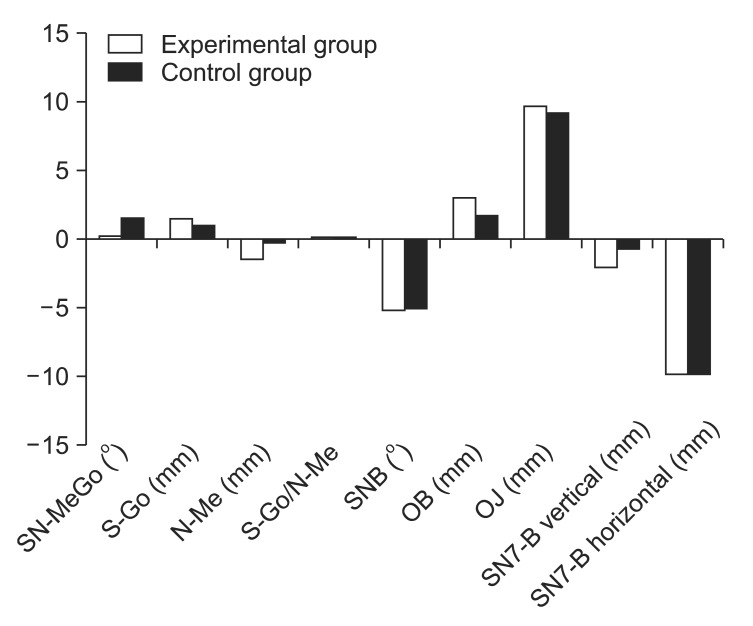
Fig. 5 Surgical movement in the experimental group and control group. There were no significant differences between the two groups (P<0.05). (SN-MeGo: mandibular plane angle, S-Go: posterior facial height, N-Me: anterior facial height, SNB: SN to NB, OB: overbite, OJ: overjet, SN7-B vertical: vertical change of B point by surgery, SN7V-B horizontal: horizontal change of B point by surgery)
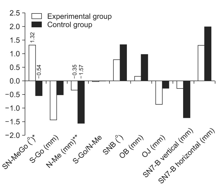
Fig. 6 Post-operative changes of parameters in the experimental group and control group six months after surgery (T2-T1). There were no significant differences between the two groups, except in mandibular plane angle (SN-MeGo) and anterior facial height (N-Me). P<0.05, *P=0.015, **P=0.029. (S-Go: posterior facial height, SNB: SN to NB, OB: overbite, OJ: overjet, SN7-B vertical: vertical change of B point by surgery, SN7V-B horizontal: horizontal change of B point by surgery)
Reference
-
1. Trauner R, Obwegeser H. The surgical correction of mandibular prognathism and retrognathia with consideration of genioplasty. I. Surgical procedures to correct mandibular prognathism and reshaping of the chin. Oral Surg Oral Med Oral Pathol. 1957; 10:677–689. PMID: 13441284.2. Dal pont G. Retromolar osteotomy for the correction of prognathism. J Oral Surg Anesth Hosp Dent Serv. 1961; 19:42–47. PMID: 13719390.3. Hunsuck EE. A modified intraoral sagittal splitting technic for correction of mandibular prognathism. J Oral Surg. 1968; 26:250–253. PMID: 5237786.4. Mehra P, Castro V, Freitas RZ, Wolford LM. Complications of the mandibular sagittal split ramus osteotomy associated with the presence or absence of third molars. J Oral Maxillofac Surg. 2001; 59:854–858. PMID: 11474434.
Article5. Moos KF, Ayoub AF. The surgical correction of dentofacial deformities, past, present and future. Egypt J Oral Maxillofac Surg. 2010; 1:2–6.6. van Merkesteyn JP, Groot RH, van Leeuwaarden R, Kroon FH. Intra-operative complications in sagittal and vertical ramus osteotomies. Int J Oral Maxillofac Surg. 1987; 16:665–670. PMID: 3125263.
Article7. Bays RA, Bouloux GF. Complications of orthognathic surgery. Oral Maxillofac Surg Clin North Am. 2003; 15:229–242. PMID: 18088677.
Article8. Falter B, Schepers S, Vrielinck L, Lambrichts I, Thijs H, Politis C. Occurrence of bad splits during sagittal split osteotomy. Oral Surg Oral Med Oral Pathol Oral Radiol Endod. 2010; 110:430–435. PMID: 20452254.
Article9. Wolford LM, Bennett MA, Rafferty CG. Modification of the mandibular ramus sagittal split osteotomy. Oral Surg Oral Med Oral Pathol. 1987; 64:146–155. PMID: 3476891.
Article10. Eggensperger N, Smolka W, Rahal A, Iizuka T. Skeletal relapse after mandibular advancement and setback in single-jaw surgery. J Oral Maxillofac Surg. 2004; 62:1486–1496. PMID: 15573348.
Article11. Eggensperger N, Smolka K, Luder J, Iizuka T. Short- and long-term skeletal relapse after mandibular advancement surgery. Int J Oral Maxillofac Surg. 2006; 35:36–42. PMID: 16344217.
Article12. Yang HJ, Hwang SJ. Postoperative stability following maxillary downward movement with Le Fort I inclined osteotomy at the lateral nasal cavity wall. J Craniomaxillofac Surg. 2012; 40:793–798. PMID: 22429608.
Article13. Mensink G, Verweij JP, Frank MD, Eelco Bergsma J, Richard van Merkesteyn JP. Bad split during bilateral sagittal split osteotomy of the mandible with separators: a retrospective study of 427 patients. Br J Oral Maxillofac Surg. 2013; 51:525–529. PMID: 23305697.
Article14. Kriwalsky MS, Maurer P, Veras RB, Eckert AW, Schubert J. Risk factors for a bad split during sagittal split osteotomy. Br J Oral Maxillofac Surg. 2008; 46:177–179. PMID: 18063456.
Article15. O'Ryan F. Complications of orthognathic surgery. Oral Maxillofac Surg Clin North Am. 1990; 2:593–601.16. Patterson AL, Bagby SK. Posterior vertical body osteotomy (PVBO): a predictable rescue procedure for proximal segment fracture during sagittal split ramus osteotomy of the mandible. J Oral Maxillofac Surg. 1999; 57:475–477. PMID: 10199504.
Article17. Tucker MR. Prevention and management of bad splits in sagittal split osteotomies. J Oral Maxillofac Surg. 2004; 62:14.
Article18. O'ryan F, Poor DB. Completing sagittal split osteotomy of the mandible after fracture of the buccal plate. J Oral Maxillofac Surg. 2004; 62:1175–1176. PMID: 15346376.19. Chrcanovic BR, Freire-Maia B. Risk factors and prevention of bad splits during sagittal split osteotomy. Oral Maxillofac Surg. 2012; 16:19–27. PMID: 21837430.
Article20. Veras RB, Kriwalsky MS, Hoffmann S, Maurer P, Schubert J. Functional and radiographic long-term results after bad split in orthognathic surgery. Int J Oral Maxillofac Surg. 2008; 37:606–611. PMID: 18515045.
Article21. Marsell R, Einhorn TA. The biology of fracture healing. Injury. 2011; 42:551–555. PMID: 21489527.
Article22. Gerstenfeld LC, Alkhiary YM, Krall EA, Nicholls FH, Stapleton SN, Fitch JL, et al. Three-dimensional reconstruction of fracture callus morphogenesis. J Histochem Cytochem. 2006; 54:1215–1228. PMID: 16864894.
Article23. LaStayo PC, Winters KM, Hardy M. Fracture healing: bone healing, fracture management, and current concepts related to the hand. J Hand Ther. 2003; 16:81–93. PMID: 12755160.
Article24. Shapiro F. Cortical bone repair. The relationship of the lacunar-canalicular system and intercellular gap junctions to the repair process. J Bone Joint Surg Am. 1988; 70:1067–1081. PMID: 3042791.
Article25. Kaderly RE. Primary bone healing. Semin Vet Med Surg (Small Anim). 1991; 6:21–25. PMID: 2038620.26. Augat P, Margevicius K, Simon J, Wolf S, Suger G, Claes L. Local tissue properties in bone healing: influence of size and stability of the osteotomy gap. J Orthop Res. 1998; 16:475–481. PMID: 9747790.
Article27. Haranal SR, Neeli AS, Suryavanshi RK, Kotrashetti SM, Naresh N. Titanium lag screw osteosynthesis in the management of mandibular fractures. Int Multidiscip Res J. 2012; 2:5–8.28. Assael LA. Evaluation of rigid internal fixation of mandible fractures performed in the teaching laboratory. J Oral Maxillofac Surg. 1993; 51:1315–1319. PMID: 8229410.
Article29. Nooh N. Stability of the mandible after bilateral sagittal split osteotomy: comparison between positioning screws and plate. Saudi Dent J. 2009; 21:123–126. PMID: 23960470.
Article30. Hammer B, Ettlin D, Rahn B, Prein J. Stabilization of the short sagittal split osteotomy: in vitro testing of different plate and screw configurations. J Craniomaxillofac Surg. 1995; 23:321–324. PMID: 8530709.
Article31. Epker BN, Fish LC. Dentofacial deformities. St. Louis: Mosby;1986. Vol. 1:p. 232–234.32. Vertenten G, Gasthuys F, Cornelissen M, Schacht E, Vlaminck L. Enhancing bone healing and regeneration: present and future perspectives in veterinary orthopaedics. Vet Comp Orthop Traumatol. 2010; 23:153–162. PMID: 20422117.
Article33. Kao ST, Scott DD. A review of bone substitutes. Oral Maxillofac Surg Clin North Am. 2007; 19:513–521. PMID: 18088902.
Article34. Hoffer MJ, Griffon DJ, Schaeffer DJ, Johnson AL, Thomas MW. Clinical applications of demineralized bone matrix: a retrospective and case-matched study of seventy-five dogs. Vet Surg. 2008; 37:639–647. PMID: 19134086.
Article35. Tiedeman JJ, Connolly JF, Strates BS, Lippiello L. Treatment of nonunion by percutaneous injection of bone marrow and demineralized bone matrix. An experimental study in dogs. Clin Orthop Relat Res. 1991; (268):294–302. PMID: 2060222.
- Full Text Links
-
- Actions
-
Cited
- CITED
-
- Close
- Share
-
- Similar articles
-
- Facial palsy after sagittal split ramus osteotomies: Case report
- Skeletal relapse pattern after sagittal split ramus osteotomy of mandibular prognathic patient
- Postoperative positional change of condyle after bilateral sagittal split ramus osteotomy associated with mandibular asymmetry
- Stability after Surgical Correction of Mandibular Prognathism Using Bilateral Sagittal Split Ramus Osteotomy with Rigid Fixation
- Treatment of osteomyelitis in the rear area of the lingula of the mandible using sagittal split ramus osteotomy: a case report
