Primary Renal Carcinoid Tumor Mimicking Non-Clear Cell Renal Cell Carcinoma: A Case Report
- Affiliations
-
- 1Department of Radiology, Keimyung University School of Medicine, Dongsan Medical Center, Daegu, Korea. kseehdr@dsmc.or.kr
- 2Department of Pathology, Keimyung University School of Medicine, Dongsan Medical Center, Daegu, Korea.
- KMID: 2327365
- DOI: http://doi.org/10.3348/jksr.2016.75.1.37
Abstract
- Carcinoid tumors are neoplasms with neuroendocrine differentiation, and they are most commonly found in the gastrointestinal and respiratory systems. Primary renal carcinoid tumor has rarely been reported. Here, we present a case of primary renal carcinoid tumor manifesting as a small but a gradually enhancing mass with calcification and a cystic component.
Figure
-
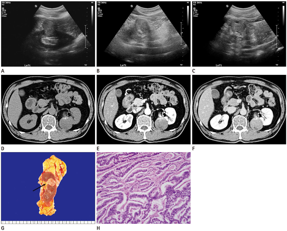
Fig. 1 A 55-year-old male with primary renal carcinoid tumor. A-C. USG findings show a slightly heterogeneous echogenic mass with focal highly echogenic foci and a cystic portion in the left renal cortex. D-F. Axial images of the dynamic abdominal CT scans demonstrate a well-circumscribed round mass (approximately 45 × 41 mm in size) with focal calcifications and a cystic component. The mass was nearly iso-attenuated (33 HU) on a pre-contrast image (D), and it showed gradual enhancement, 53 HU on the corticomedullary phase image (E), and 83 HU on the excretory phase image (F). G. Gross specimen of the mass shows a well circumscribed mass, measuring 4.0 × 3.9 × 2.5 cm in size. The mass is located in the upper pole cortex of the left kidney and shows direct invasion into perinephric tissue (arrow). H. The tumor cells exhibit a characteristic ribbon-like pattern (hematoxylin and eosin stain, × 200). CT = computed tomography, HU = Hounsfield unit, USG = ultrasonography
Reference
-
1. Modlin IM, Sandor A. An analysis of 8305 cases of carcinoid tumors. Cancer. 1997; 79:813–829.2. Romero FR, Rais-Bahrami S, Permpongkosol S, Fine SW, Kohanim S, Jarrett TW. Primary carcinoid tumors of the kidney. J Urol. 2006; 176(6 Pt 1):2359–2366.3. Kim JM, Lee JH. Carcinoid tumor arising from horseshoe kidney: report of two cases. J Korean Radiol Soc. 2001; 44:509–512.4. Murali R, Kneale K, Lalak N, Delprado W. Carcinoid tumors of the urinary tract and prostate. Arch Pathol Lab Med. 2006; 130:1693–1706.5. Moulopoulos A, DuBrow R, David C, Dimopoulos MA. Primary renal carcinoid: computed tomography, ultrasound, and angiographic findings. J Comput Assist Tomogr. 1991; 15:323–325.6. Shurtleff BT, Shvarts O, Rajfer J. Carcinoid tumor of the kidney: case report and review of the literature. Rev Urol. 2005; 7:229–233.7. Yoon JH. Primary renal carcinoid tumor: a rare cystic renal neoplasm. World J Radiol. 2013; 5:328–333.8. Millet I, Doyon FC, Hoa D, Thuret R, Merigeaud S, Serre I, et al. Characterization of small solid renal lesions: can benign and malignant tumors be differentiated with CT? AJR Am J Roentgenol. 2011; 197:887–896.9. Kim JK, Kim TK, Ahn HJ, Kim CS, Kim KR, Cho KS. Differentiation of subtypes of renal cell carcinoma on helical CT scans. AJR Am J Roentgenol. 2002; 178:1499–1506.10. Fujimoto H, Wakao F, Moriyama N, Tobisu K, Sakamoto M, Kakizoe T. Alveolar architecture of clear cell renal carcinomas (< or = 5.0 cm) show high attenuation on dynamic CT scanning. Jpn J Clin Oncol. 1999; 29:198–203.
- Full Text Links
-
- Actions
-
Cited
- CITED
-
- Close
- Share
-
- Similar articles
-
- A case of renal transitional cell carcinoma associated with synchronous contralateral renal cell carcinoma
- A Case of Papillary Type of Renal Cell Carcinoma after Renal Injury in a Child
- Carcinoid Tumor in Horseshoe Kidney
- A Case of Metastatic Renal Cell Carcinoma to the Gallbladder
- Differentiation of Chromophobe Renal Cell Carcinoma and Clear Cell Renal Cell Carcinoma by Using Helical CT
