Retroperitoneal Leiomyoma of the Uterus Mimicking Sarcoma in Perimenopausal Woman: Case Report
- Affiliations
-
- 1Department of Surgery, Soonchunhyang University College of Medicine, Bucheon, Korea. gwsdlove@schmc.ac.kr
- KMID: 2325479
- DOI: http://doi.org/10.6118/jmm.2014.20.3.133
Abstract
- Leiomyomas are very common benign tumors in the uterus and it is rare condition to present the retroperitoneal leiomyoma. The author reported a 48-year-old female patient who presented right pelvic mass with urinary incontinence and lower abdominal discomfort. Based on the preoperative imaging, provisional diagnosis was mesenchymal sarcoma. In the intraoperative findings, huge mass abutting to the uterus was observed in retroperitoneal space beneath the right broad ligament. After the exposure the retroperitoneal space, we encountered the well-demarcated tumor measuring 8 x 6 cm in diameter and this tumor attached the right surface of the uterus with fibrotic tissue. Pathologic findings demonstrated retroperitoneal uterine leiomyoma.
Keyword
MeSH Terms
Figure
-
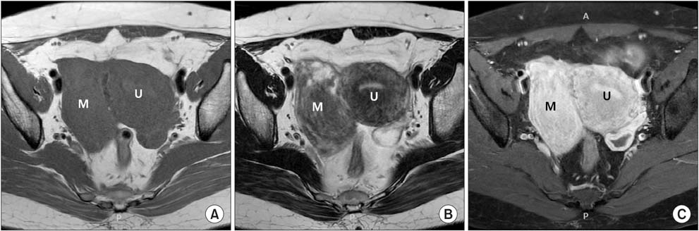
Fig. 1 Pelvic magnetic resonance imaging showed 8-cm sized mass separating from uterus. This mass had the low signal intensity on T1-weighted images with enhancement and heterogeneous, high signal intensity on T2-weighted images. (A) Axial, T1-weighted image, (B) axial, T2-weighted image, (C) axial, enhanced T1-weighted image. U: uterus, M: retroperitoneal mass.

Fig. 2 The operative findings. (A) The uterus was normal size with smooth contour and there was no abnormal finding on both ovaries and fallopian tubes grossly. (B) The retroperitoneal tumor of size 8 × 6 cm was located beneath the right broad ligament of uterus. (C) After dissection from the right iliac vessels and the right surface of the uterus, the retroperitoneal tumor was excised completely. U: uterus, B: broad ligament, M: retroperitoneal mass.
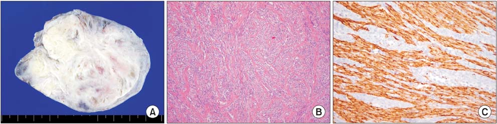
Fig. 3 The macroscopic and microscopic findings. (A) Grossly, it is well circumscribed, and the cut surface shows gray-white whirling appearance. (B) The tumor consists of intersecting fascicles of slender-tapered spindle cells (H & E, ×100). (C) Immunohistochemical stain for desmin revealed positivity in the tumor cells, consistent with smooth muscle tumor (×200).
Reference
-
1. Albert PS, Sinatra T, Nagamatsu GR. Retroperitoneal leiomyoma presenting as prostatic mass. Urology. 1974; 3:607–609.2. Dalen T, Coebergh JW, Casparie MK, Gimbrère CH, Hoekstra HJ, Van Geel BN, et al. Soft tissue sarcoma: the predominant primary malignancy in the retroperitoneum. Sarcoma. 2001; 5:5–8.3. Erzen D, Sencar M, Novak J. Retroperitoneal sarcoma: 25 years of experience with aggressive surgical treatment at the Institute of Oncology, Ljubljana. J Surg Oncol. 2005; 91:1–9.4. Buttram VC Jr, Reiter RC. Uterine leiomyomata: etiology, symptomatology, and management. Fertil Steril. 1981; 36:433–445.5. Poliquin V, Victory R, Vilos GA. Epidemiology, presentation, and management of retroperitoneal leiomyomata: systematic literature review and case report. J Minim Invasive Gynecol. 2008; 15:152–160.6. Mahendru R, Gaba G, Yadav S, Gaba G, Gupta C. A rare case of retroperitoneal leiomyoma. Case Rep Surg. 2012; 2012:425280.7. Fasih N, Prasad Shanbhogue AK, Macdonald DB, Fraser-Hill MA, Papadatos D, Kielar AZ, et al. Leiomyomas beyond the uterus: unusual locations, rare manifestations. Radiographics. 2008; 28:1931–1948.8. Kho KA, Nezhat C. Parasitic myomas. Obstet Gynecol. 2009; 114:611–615.9. Moon HS, Koo JS, Park SH, Park GS, Choi JG, Kim SG. Parasitic leiomyoma in the abdominal wall after laparoscopic myomectomy. Fertil Steril. 2008; 90:1201.e1–1201.e2.10. Van Roggen JF, Hogendoorn PC. Soft tissue tumours of the retroperitoneum. Sarcoma. 2000; 4:17–26.11. Kondo W, Botchorishvili R, Desvignes F, Mage G. Laparoscopic management of a pelvic retroperitoneal leiomyomacase report. Gynecol Surg. 2011; 8:247–251.
- Full Text Links
-
- Actions
-
Cited
- CITED
-
- Close
- Share
-
- Similar articles
-
- A case of Huge Isolated Retroperitoneal Leiomyoma
- Retroperitoneal Leiomyoma With Exophytic Growing into the Lesser Sac: A Case Report
- Exophytic Lipoleiomyoma of the Uterus Mimicking Ovarian Teratoma: A Case Report
- A case of liposarcoma within a leiomyoma
- Retroperitoneal Erdheim-Chester disease without skeletal bone involvement mimicking uterine sarcoma with multiple organ involvement
