Ganglion and Synovial Cyst on the Posterior Longitudinal Ligament: Case Report
- Affiliations
-
- 1Department of Orthopedic Surgery, Han-il Hospital. pooljang105@hanmail.net
Abstract
- Ganglion cyst and synovial cyst are rarely found in the lumbar spinal canal. Synovial cysts usually arise from degenerative apophyseal joints, and ganglion cysts arise from articular facet joints. We present here two cases: one ganglion cyst that arose from the posterior longitudinal ligament at the L2-3 intervertebral space level, and a synovial cyst that also arose from the posterior longitudinal ligament at the level of the L4 body. Both patients progressively developed low back pain with radiating pain. The lesions were detected by magnetic resonance imaging in both cases. The dural sac was displaced by the cysts in both cases. Relief of symptoms was achieved by decompressive laminectomy and excision of the ganglion cyst, and excision of the synovial cyst and PLIF at L3-4. Because these lesions have not been reported on before, we report here on 2 cases with synovial and ganglion cyst from the posterior longitudinal ligament at lumbar spinal level.
Keyword
MeSH Terms
Figure
-

Fig. 1. MR images show ganglion cyst located on the L2 level of posterior longitudinal ligament. (A) sagital T1 & T2-weighted image: 1.2× 0.8× 0.7 cm sized mass with low signal in T1, high signal in T2 images (B) axial T1 & T2-weighted image: 1.2× 0.8× 0.7 cm sized mass with low sinal in T1, high signal in T2 images (C) excised ganglion cyst (D) photomicrograph of section of ganglion cyst, which shows capsule but no surrounding fibrous tissue.
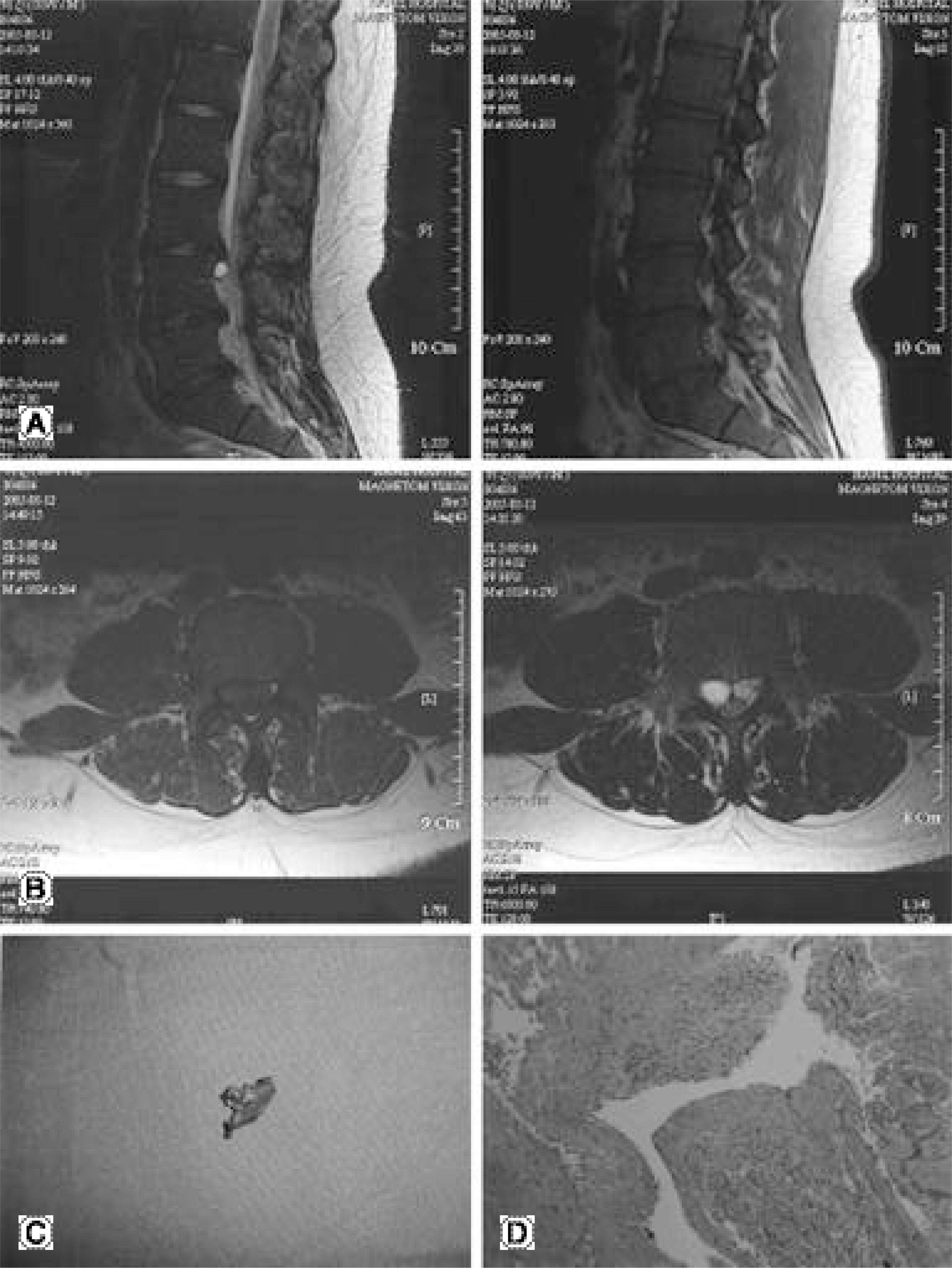
Fig. 2. MR images show synovial cyst located on the L4 level of posterior longitudinal ligament. (A) sagital T1 & T2-weighted image: 1.4× 1.2× 0.9 cm and 0.8× 0.7× 0.6 cm sized masses with low signal in T1, high signal in T2 images (B) axial T1 & T2-weighted image: 1.4× 1.2× 0.9 cm and 0.8x0.7x0.6cm sized masses with low signal in T1, high signal in T2 images (C) excised synovial cyst after aspiration during operation (D) photomicrograph of section of synovial cyst, which shows mesothelial cells surrounding the cystic materials.
Reference
-
1). Abdullah AF, Chambers RW, Daut DP. Lumbar nerve root compression by synovial cysts of the ligamentum flavum: Report of four cases. J. Neurosurg. 60:617–620. 1984.2). Awwad EE, Martin DS, Smith KR Jr, Bucholz RD. MR imaging of lumbar juxtaarticular cysts. J Comput Assist Tomogr. 14:415–417. 1990.
Article3). Bhushan C, Hodges FJ 3rd, Wityk JJ. S y n o v i a l cysts(ganglion) of the lumbar spine simulating extradural mass. Neuroradiology. 18:263–268. 1979.4). Herrington JL Jr., Edwards LW. Ganglion cysts arising in unusual locations. Ann Surg. 142:900–903. 1955.
Article5). Liu SS, Williams KD, Drayer BP, Spetzler RF, Sonntag VK. synovial cysts of the lumbaosacral spine: diagnosis by MR imaging. AJR Am J Roentgenol. 154:163–166. 1990.6). Azzam CJ. Midline lumbar ganglion/synovial cyst mimic -king an epidural tumor: Case report and review of patho -genesis. Neurosurgery. 23:232–234. 1988.7). Kjerulf TD, Terry DW Jr, Boubelik RJ. Lumbar synovial or ganglion cysts. Neurosurgery. 19:415–420. 1986.
Article8). Pendleton B, Carl B, Pollay M. Spinal extradural benign synovial or ganglion cyst: Case report and review of the literature. Neurosurgery. 13:322–326. 1983.
Article9). Chiang KS, Lee YY, Mawad ME. MR of intraspinal synovial cyst: Rim enhancement with gadopentetate dimeglu -mine. AJR Am J Roentgenol. 157:416. 1991.10). Kao CC, Uihlein A, Bickel WH, Soule EH. Lum b ar intraspinal extradural ganglion cyst. J Neurosurg. 29:168–172. 1968.11). Jackson DE Jr, Atlas SW, Mani JR, Norman D. Intraspinal synovial cysts: MR imaging. Radiology. 170:527–530. 1989.
Article12). Yuh WTC et al: Intraspinal synovial cysts. Magn etic resonance evaluation. Spine. 16:740–745. 1991.
- Full Text Links
-
- Actions
-
Cited
- CITED
-
- Close
- Share
-
- Similar articles
-
- Ganglion Cyst of the Posterior Cruciate Ligament: A Case Report
- Ganglion Cyst of the Posterior Longitudinal Ligament Causing Lumbar Radiculopathy
- Intraarticular Ganglion Arising from the Posterior Cruciate Ligament
- Lumbar intraspinal Extradural Cysts: 2 cases report
- Symptomatic Posterior Cruciate Ganglion Cyst Causing Impingement between Posterior Root of the Medial Meniscus and Anterior to the Posterior Cruciate Ligament
