A morphologic evaluation of defects created by a piezoelectric ultrasonic scaler on casting gold alloy
- Affiliations
-
- 1Department of Periodontics, Asan Medical Center, Seoul, Korea. periolee@amc.seoul.kr
- 2Department of Dentistry, College of Medicine, Ulsan University, Ulsan, Korea.
Abstract
- PURPOSE
In this study we evaluated the morphologic aspects of defects created by a piezoelectric ultrasonic scaler with scaler tip on casting gold alloy using scanning electron microscope (SEM) images and defect surface profiles. METHODS: 54 blocks of type III casting gold alloy (Firmilay, Jellenko Inc, CA, USA) were scaled by a piezoelectric ultrasonic scaler (P-MAX, Satelec, France) with scaler tip (No. 1 tip) on a sledge device. 2-dimensional profiles of defects on all samples were investigated by a surface profilometer (a-Step 500, KLA-Tencor, CA, USA). The selected working parameters were lateral force (0.5 N, 1.0 N, 2.0 N), mode (P mode, S mode), and power setting (2, 4, 8). SEM images were obtained. Defect surface profiles were made on Microsoft Excel program using data obtained by a surface profilometer. RESULTS: Among P mode samples, there were similarities on defect surface profiles and SEM images regardless of lateral force. The defects created in P mode were narrow and shallow although the depth and the width increased as power setting changed low (2) to high (8). In P mode samples, the defect depth was the greatest when lateral force of 0.5 N was applied. However all the depths were smaller than 1 m. SEM images of Lateral force of 0.5 N, S mode, power setting 2 and 4 were similar to that of P mode, but the other SEM images of S mode showed discernible changes. Defect depth of S mode samples was the greatest when lateral force of 1.0 N was applied. CONCLUSIONS: Within the limitations of this study, it can be concoluded that removing capability of piezoelectric scaler with scaler tip becomes maximized as power level becomes higher but the capability is restricted when excessive lateral force is applied on scaler tip.
Keyword
Figure
-
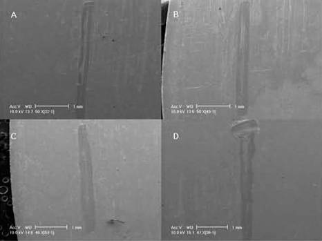
Figure 1 Scanning electon microscope image of defects. A) lateral force 0.5 N, P mode, power setting 2, magnification ×50, B) lateral force 1.0 N, P mode, power setting 4, magnification ×50, C) lateral force 2.0 N, P mode, power setting 4, magnification ×48, D) lateral force 0.5 N, S mode, power setting 4, magnification ×47.
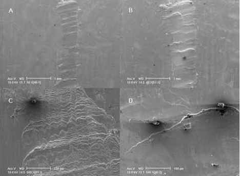
Figure 2 Scanning electron microscope image of defects. A) lateral force 1.0 N, S mode, power setting 4, magnification ×50, B) lateral force 2.0 N, S mode, power setting 8, magnification ×49, C) lateral force 0.5 N, S mode, power setting 8, magnification X500, D) lateral force 1.0 N, S mode, power setting 4, magnification X500.
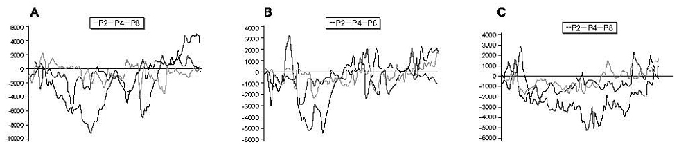
Figure 3 Surface profile of defects created at P mode. A) lateral force 0.5 N, B) lateral force 1.0 N, C) lateral force 2.0 N. In the box, P2, P4 and P8 mean P mode power setting 2, P mode power setting 4 and P mode power setting 8, respectively.
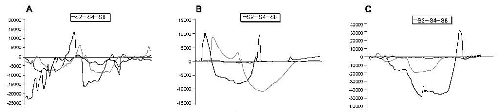
Figure 4 Surface profile of defects created at S mode. A) lateral force 0.5 N, B) lateral force 1.0 N, C) lateral force 2.0 N. In the box, S2, S4 and S8 mean S mode power setting 2, S mode power setting 4 and S mode power setting 8, respectively.
Reference
-
1. Socransky SS, Haffajee AD. Dental biofilms: difficult therapeutic targets. Periodontol 2000. 2002. 28:12–55.
Article2. Breininger DR, O'Leary TJ, Blumenshine RV. Comparative effectiveness of ultrasonic and hand scaling for the removal of subgingival plaque and calculus. J Periodontol. 1987. 58:9–18.
Article3. Gellin RG, Miller MC, Javed T, Engler WO, Mishkin DJ. The effectiveness of the Titan-S sonic scaler versus curettes in the removal of subgingival calculus: a human surgical evaluation. J Periodontol. 1986. 57:672–680.
Article4. Zappa U, Smith B, Simona C, et al. Root substance removal by scaling and root planning. J Periodontol. 1991. 62:750–754.5. Kerry GJ. Roughness of root surfaces after use of ultrasonic instruments and hand curettes. J Periodontol. 1967. 38:340–346.
Article6. Wilkinson RF, Maybury JE. Scanning electron microscopy of the root surface following instrumentation. J Periodontol. 1973. 44:559–563.
Article7. Zappa U, Cadosch J, Simona C, Graf H, Case D. In vivo scaling and root planing forces. J Periodontol. 1991. 62:335–340.
Article8. Ruppert M, Cadosch J, Guindy J, Case D, Zappa U. In vivo ultrasonic debridement forces in bicuspids: A pilot study. J Periodontol. 2002. 73:418–422.9. Drisko CH. Root instrumentation: power-driven versus manual scalers, which one? Dent Clin North Am. 1998. 42:229–244.10. Flemmig TF, Petersilka GJ, Mehl A, Hickel R, Klaiber B. The effect of working parameters on root substance removal using a piezoelectric ultrasonic scaler in vitro. J Clin Periodontol. 1998. 25:158–163.
Article11. Lee YK. Root surface roughness following mechanical instrumentation in vivo and in vitro SEM study. J Korean Acad Periodontol. 1998. 28:823–828.
Article12. Lee YK. The effect of a piezoelectric ultrasonic scaler with curette tip on root substitute removal in Vitro. J Korean Acad Periodontol. 2000. 30:429–441.
Article13. Lee YK. The effect of a piezoelectric ultrasonic scaler with curette tip on casting gold removal in vitro. J Korean Acad Periodontol. 2001. 31:531–542.
Article14. Cha KB, Kim WK, Lee YK, Kim YS. The effect of working parameters on removal of casting gold alloy using a piezoelectric ultrasonic scaler with scaler tip in vitro. J Korean Acad Periodontol. 2009. 39:139–148.
Article
- Full Text Links
-
- Actions
-
Cited
- CITED
-
- Close
- Share
-
- Similar articles
-
- The effect of working parameters on removal of casting gold alloy using a piezoelectric ultrasonic scaler with scaler tip in vitro
- The Effect of a Piezoelectric Ultrasonic Scaler with Curette Tip on Casting Gold Removal in Vitro
- A STUDY OF INTERFACE AND CORROSION BEHAVIOR BETWEEN IMPLANT ABUTMENT AND CASTING GOLD ALLOY
- The Effect of a Piezoelectric Ultrasonic Scaler with Curette Tip on Root Substitute Removal in Vitro
- Evaluation of Pain Reduction and Clinical Efficacy of Feedback-Controlled Ultrasonic Scaler
