Pulmonary Foreign Body Granulomatosis in Dental Technician
- Affiliations
-
- 1Department of Internal Medicine, Hanyang University College of Medicine, Seoul, Korea. drterry@hanyang.ac.kr
- 2Department of Pathology, Hanyang University College of Medicine, Seoul, Korea.
- KMID: 2320725
- DOI: http://doi.org/10.4046/trd.2015.78.4.445
Abstract
- Occupational lung diseases are caused by several toxic substances including heavy metals; however, the exact pathologic mechanisms remain unknown. In the workplace, dental technicians are often exposed to heavy metals such as cobalt, nickel, or beryllium and occasionally develop occupational lung diseases. We described a case of occupational lung disease in a patient who was employed as a dental technician for over a decade. A 31-year-old, non-smoking woman presented with productive cough and shortness of breath of several weeks duration. Chest computed tomography revealed a large number of scattered, bilateral small pulmonary nodules throughout the lung field, and multiple mediastinal lymph nodes enlargement. Percutaneous needle biopsy showed multifocal small granulomas with foreign body type giant cells suggestive of heavy metals inhalation. The patient's condition improved on simple avoidance strategy for several months. This case highlighted the importance of proper workplace safety.
Keyword
MeSH Terms
Figure
-
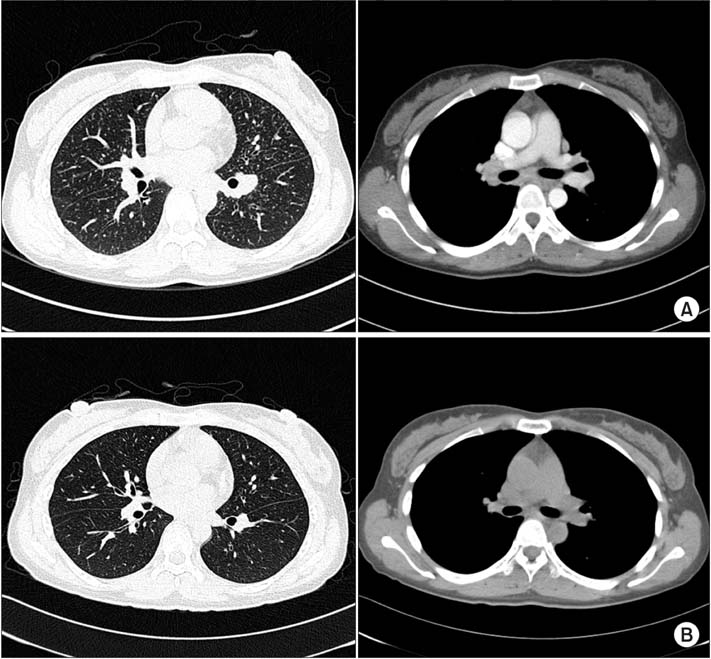
Figure 1 Chest computed tomography (CT) on admission (A) and at 3 months after suitable precaution (B). (A) Chest CT on admission showed multiple small nodules in both lower lung fields. Both hilar and mediastinal lymph nodes enlargement are also seen. (B) After 3 months, multiple small nodules are reduced in size and number. Enlargement of both-hilar and mediastinal lymph nodes are absent.
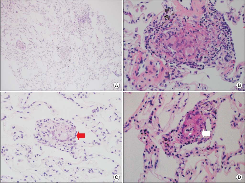
Figure 2 Histologically, the lung biopsy reveals multifocal small and discrete non-caseating epithelioid granulomas containing foreign body-type giant cells. (A) Multifocal granulomas shown in a low power field (H&E stain, ×100). (B) Representative non-caseating epithelioid granulomas containing giant cells engulfing foreign bodies in a high power field (H&E stain, ×400). (C) Typical granuloma containing giant cells engulfing refractile foreign bodies (arrow) (H&E stain, ×400). (D) Typical granulomas containing foreign body-type giant cells engulfing refractile foreign bodies (arrow), which are conspicuous under polarized light microscopy (H&E stain, ×200).
Reference
-
1. Brancaleone P, Weynand B, De Vuyst P, Stanescu D, Pieters T. Lung granulomatosis in a dental technician. Am J Ind Med. 1998; 34:628–631.2. Park SY, Kim HR, Song J. Workers' compensation for occupational respiratory diseases. J Korean Med Sci. 2014; 29:Suppl. S47–S51.3. Ozdemir Dogan D, Ozdemir AK, Polat NT, Dal U, Gumus C, Akkurt I. Prevalence of respiratory abnormalities and pneumoconiosis in dental laboratory technicians. Tuberk Toraks. 2010; 58:135–141.4. Torbica N, Krstev S. World at work: dental laboratory technicians. Occup Environ Med. 2006; 63:145–148.5. Froudarakis ME, Voloudaki A, Bouros D, Drakonakis G, Hatzakis K, Siafakas NM. Pneumoconiosis among Cretan dental technicians. Respiration. 1999; 66:338–342.6. Thorette C, Grigoriu B, Canut E, Sobaszek A, Tonnel AB, Tillie-Leblond I. Pulmonary disease in dental laboratory technicians. Rev Mal Respir. 2006; 23:Suppl 2. 4S7–4S16.7. Abakay A, Atilgan S, Abakay O, Atalay Y, Guven S, Yaman F, et al. Frequency of respiratory function disorders among dental laboratory technicians working under conditions of high dust concentration. Eur Rev Med Pharmacol Sci. 2013; 17:809–814.8. Kartaloglu Z, Ilvan A, Aydilek R, Cerrahoglu K, Tahaoglu K, Baloglu H, et al. Dental technician's pneumoconiosis: mineralogical analysis of two cases. Yonsei Med J. 2003; 44:169–173.9. Cimrin A, Komus N, Karaman C, Tertemiz KC. Pneumoconiosis and work-related health complaints in Turkish dental laboratory workers. Tuberk Toraks. 2009; 57:282–288.10. Choudat D. Occupational lung diseases among dental technicians. Tuber Lung Dis. 1994; 75:99–104.11. Choudat D, Triem S, Weill B, Vicrey C, Ameille J, Brochard P, et al. Respiratory symptoms, lung function, and pneumoconiosis among self employed dental technicians. Br J Ind Med. 1993; 50:443–449.12. Hu SW, Lin YY, Wu TC, Hong CC, Chan CC, Lung SC. Workplace air quality and lung function among dental laboratory technicians. Am J Ind Med. 2006; 49:85–92.
- Full Text Links
-
- Actions
-
Cited
- CITED
-
- Close
- Share
-
- Similar articles
-
- Management of Gastrointestinal Foreign Body Ingested during Dental Procedure
- Prevention and management of foreign body ingestion and aspiration during the dental treatment
- Primary Lymphomatoid Granulomatosis in the Frontal Lobe: Case Report
- Management of Foreign Body Ingestion during Implant Surgery: A Case Report
- Dental Hygienists' Awareness of Medical Technician Jurisprudence
