Tuberc Respir Dis.
2015 Oct;78(4):408-411. 10.4046/trd.2015.78.4.408.
Successful Treatment of a Large Pulmonary Arteriovenous Malformation by Repeated Coil Embolization
- Affiliations
-
- 1Division of Pulmonary and Critical Care Medicine, Department of Internal Medicine, Seoul National University Hospital, Seoul, Korea. mdyspark@gmail.com
- KMID: 2320716
- DOI: http://doi.org/10.4046/trd.2015.78.4.408
Abstract
- Pulmonary arteriovenous malformations (AVMs) are caused by abnormal vascular communications between the pulmonary arteries and pulmonary veins, which lead to the blood bypassing the normal pulmonary capillary beds. Pulmonary AVMs result in right-to-left shunts, resulting in hypoxemia, cyanosis, and dyspnea. Clinical signs and symptoms vary depending on the size, number, and flow of the AVMs. Transcatheter embolization is the treatment of choice for pulmonary AVMs. However, this method can fail if the AVM is large or has multiple complex feeding arteries. Surgical resection is necessary in those kind of cases. Here, we report the case of a patient with a 6-cm pulmonary AVM with multiple feeding arteries that was successfully treated by repeated coil embolization without surgery.
MeSH Terms
Figure
-
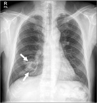
Figure 1 Initial chest radiograph showing a mass-like opacity in the right lower lung field.

Figure 2 (A-D) Chest computed tomography showing a huge entangled enhanced lesion in the right middle lobe, which is consistent with pulmonary arteriovenous malformation (arrows).

Figure 3 Angiography visualizing a large pulmonary arteriovenous malformation with multiple complex feeding arteries (A). Transcatheter embolization was performed twice (B, C), and post-embolization chest radiography revealed radioopaque embolization materials (D).
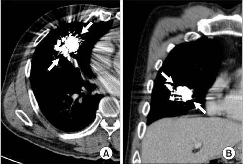
Figure 4 (A, B) A 1-year follow-up computed tomography showing no evidence of recanalization, although the evaluation was limited due to the presence of artifacts (arrows).
Reference
-
1. Shovlin CL. Pulmonary arteriovenous malformations. Am J Respir Crit Care Med. 2014; 190:1217–1228.2. Khurshid I, Downie GH. Pulmonary arteriovenous malformation. Postgrad Med J. 2002; 78:191–197.3. Cartin-Ceba R, Swanson KL, Krowka MJ. Pulmonary arteriovenous malformations. Chest. 2013; 144:1033–1044.4. Gossage JR, Kanj G. Pulmonary arteriovenous malformations: a state of the art review. Am J Respir Crit Care Med. 1998; 158:643–661.5. Lee DW, White RI Jr, Egglin TK, Pollak JS, Fayad PB, Wirth JA, et al. Embolotherapy of large pulmonary arteriovenous malformations: long-term results. Ann Thorac Surg. 1997; 64:930–939.6. Kim HJ, Lee JS, Oh YM, Shim TS, Lim CM, Koh YS, et al. Clinical characteristics of pulmonary arteriovenous malformations in Koreans. Respirology. 2015; 20:155–159.7. Iqbal M, Rossoff LJ, Steinberg HN, Marzouk KA, Siegel DN. Pulmonary arteriovenous malformations: a clinical review. Postgrad Med J. 2000; 76:390–394.8. Hsu CC, Kwan GN, Thompson SA, Evans-Barns H, van Driel ML. Embolisation for pulmonary arteriovenous malformation. Cochrane Database Syst Rev. 2015; 1:CD008017.9. Milic A, Chan RP, Cohen JH, Faughnan ME. Reperfusion of pulmonary arteriovenous malformations after embolotherapy. J Vasc Interv Radiol. 2005; 16:1675–1683.10. Hart JL, Aldin Z, Braude P, Shovlin CL, Jackson J. Embolization of pulmonary arteriovenous malformations using the Amplatzer vascular plug: successful treatment of 69 consecutive patients. Eur Radiol. 2010; 20:2663–2670.11. Kucukay F, Ozdemir M, Senol E, Okten S, Ereren M, Karan A. Large pulmonary arteriovenous malformations: long-term results of embolization with AMPLATZER vascular plugs. J Vasc Interv Radiol. 2014; 25:1327–1332.
- Full Text Links
-
- Actions
-
Cited
- CITED
-
- Close
- Share
-
- Similar articles
-
- A Case of Multiple Pulmonary Arteriovenous Malformation Treated with Coil Embolization
- A Case of Transcatheter Coil Embolization of Diffuse Pulmonary Arteriovenous Malformation
- A Case of Percutaneous Transcatheter Coil Embolization for Congenital Coronary Arteriovenous Fistula
- A Case of Embolotherapy of Diffuse Pulmonary Arteriovenous Malformation Using Amplatzer Vascular Plugs
- A Case of Giant Aneurysm of Coronary Arteriovenous Fistula Treated by Percutaneous Deployment of Embolization Coil
