Spontaneous Pulmonary Hematoma with No Underlying Causes: A Case Report
- Affiliations
-
- 1Department of Internal Medicine, Konkuk University Chungju Hospital, Chungju, Korea.
- 2Division of Pulmonary and Critical Care Medicine, Department of Internal Medicine, Konkuk University Chungju Hospital, Chungju, Korea.
- 3Department of Thoracic and Cardiovascular Surgery, Konkuk University Chungju Hospital, Chungju, Korea.
- 4Division of Allergy and Pulmonology, Department of Internal Medicine, Konkuk University Chungju Hospital, Chungju, Korea. ggulcha2000@naver.com
- KMID: 2320707
- DOI: http://doi.org/10.4046/trd.2015.78.4.363
Abstract
- A 57-year-old male patient was admitted to our center because of a cystic mass on the lower portion of the right major fissure that was found incidentally by chest X-ray. He did not have a history of trauma or anticoagulant use. The lesion was removed by video-assisted thoracoscopic surgery. Pathological examination revealed an organizing pulmonary hematoma without any complications, and a follow-up chest X-ray after 1 year showed no recurrence.
Keyword
MeSH Terms
Figure
-

Figure 1 Chest X-rays shows a well-defined round homogenous opacity in the right lower lung field. RPA: posterior anterior view.
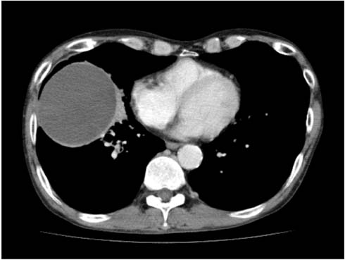
Figure 2 Chest computed tomography scanning reveals a 9.0×7.9-cm-sized well-defined cystic mass in the upper and lower lobes of the right lung.
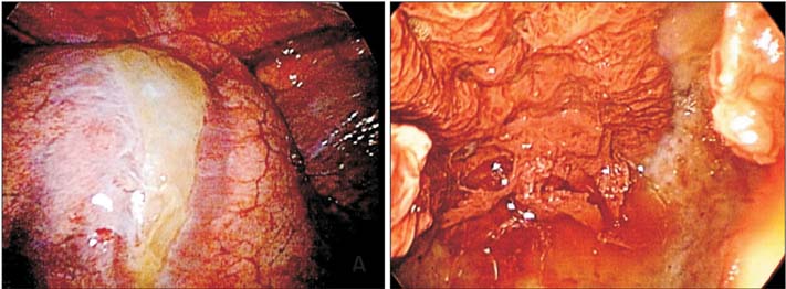
Figure 3 (A, B) Surgical findings demonstrates a large cystic mass with a thick yellowish capsule in the right upper and lower lobes, containing a large amount of dark brown muddy material.
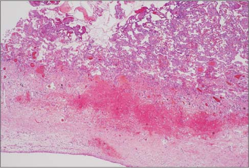
Figure 4 Histopathological findings shows dense fibrous tissue containing fragments of blood clots separating the lung parenchyma (H&E stain, ×40).
Cited by 1 articles
-
Pulmonary Comorbidities of Lung Emphysema
Hye Rim Park, Young Tong Kim, Sung Shick Jou, Chan Ho Park
J Korean Soc Radiol. 2018;79(3):139-151. doi: 10.3348/jksr.2018.79.3.139.
Reference
-
1. Trinkle JK, Richardson JD, Franz JL, Grover FL, Arom KV, Holmstrom FM. Management of flail chest without mechanical ventilation. Ann Thorac Surg. 1975; 19:355–363.2. Mathai M, Byrd RP Jr, Roy TM. The posttraumatic pulmonary mass. J Tenn Med Assoc. 1996; 89:41–42.3. Chakraborty AK, Dreisin RB. Pulmonary hematoma secondary to anticoagulant therapy. Ann Intern Med. 1982; 96:67–69.4. Kaira K, Takei Y, Matsuura M, Saito R. Pulmonary hematoma resulting from anticoagulant therapy. AJR Am J Roentgenol. 2003; 180:1740.5. Specht EE. Pulmonary hematoma. Am J Dis Child. 1966; 111:559–563.6. Williams JR. The vanishing lung tumor: pulmonary hematoma. Am J Roentgenol Radium Ther Nucl Med. 1959; 81:296–302.7. Luna MA, Leary WV, Jing BS. Pulmonary hematoma associated with thrombocytopenia. Chest. 1970; 57:487–489.8. Matsushita A, Takayanagi N, Ishiguro T, Harasawa K, Tsuchiya N, Yoneda K, et al. A case of Ehlers-Danlos syndrome suspected from pulmonary hematoma due to disruption of the lung. Nihon Kokyuki Gakkai Zasshi. 2009; 47:704–710.9. Joynt GH, Jaffe F. Solitary pulmonary hematoma. J Thorac Cardiovasc Surg. 1962; 43:291–302.10. Jay SJ, Johanson WG Jr. Massive intrapulmonary hemorrhage: an uncommon complication of bullous emphysema. Am Rev Respir Dis. 1974; 110:497–501.
- Full Text Links
-
- Actions
-
Cited
- CITED
-
- Close
- Share
-
- Similar articles
-
- Erratum: Spontaneous Pulmonary Hematoma with No Underlying Causes: A Case Report
- A Case of Spontaneous Cervical Hematoma caused by Parathyroid Adenoma
- Spontaneous Ruptured Subcapsular Liver Hematoma Associated with Pregnancy
- Spontaneous intracranial epidural hematoma following aortic valve replacement: A case report
- A Case of Spontaneous Thoracic Epidural Hematoma
