A Case of Multi-Organ Macronodular Tuberculosis
- Affiliations
-
- 1Department of Internal Medicine, Kangwon National University School of Medicine, Chuncheon, Korea. ssunimd@kangwon.ac.kr
Abstract
- A 37 year old female presented with epigastric pain and weight loss over a period of 3 months. Her abdominal CT finding showed a 4.5 cm size hepatic mass and 4.3 cm size pancreatic head mass with multiple macronodules in the liver. At the same time, her chest CT revealed a 5 cm size necrotic mass in the left lower lobe of the lung with multiple bilateral pulmonary nodules. We diagnosed these lesions as tuberculosis through multiple biopsies. She was treated with anti-tuberculous medication. After taking the medications, her symptoms were improved. Twelve months later, imaging studies indicated an improvement in the patient's health. Here we report a case report of multi-organ macronodular tuberculosis in lung, liver and pancreas.
MeSH Terms
Figure
-
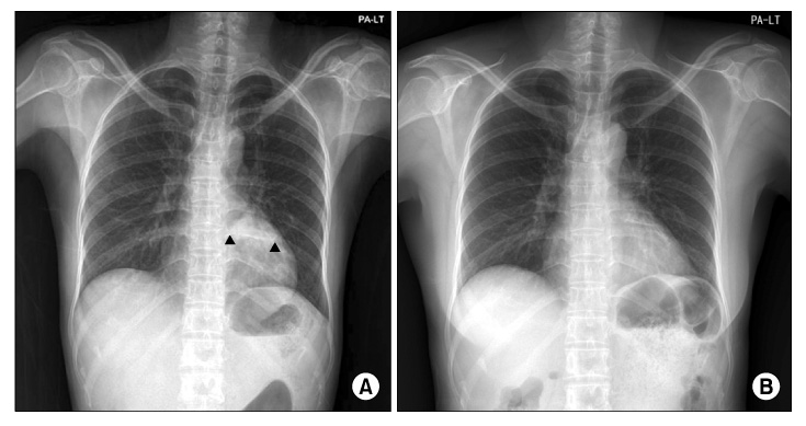
Figure 1 Simple chest PA showed an approximately 5 cm size consolidation in the retrocardiac area (A, black arrow heads) and nearly complete resolution after treatment (B).
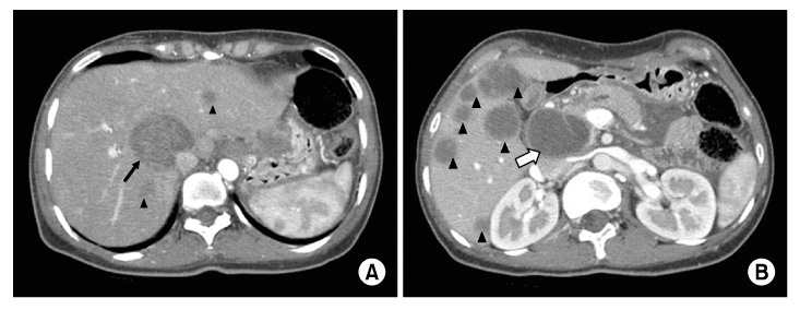
Figure 2 (A) Abdominal contrast enhanced CT images showed an approximately 4.5 cm size mass (black arrow) in the central liver. (B) Multiple variable size low density masses (black arrow heads) in the liver and a 4.5 cm size mass in the head of the pancreas (white arrow). CT: computed tomography.
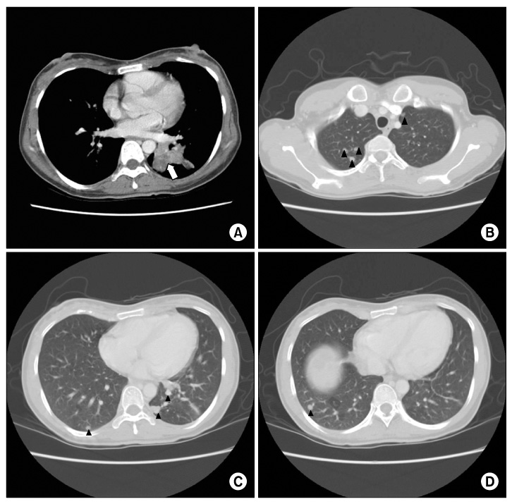
Figure 3 Chest CT images revealed a 5 cm size irregular necrotic mass (white arrow) in LLL (A) and multiple nodules (black arrow heads) in both lungs (B~D). CT: computed tomography; LLL: left lower lobe.

Figure 4 Microscopic findings of the lesion. (A) Needle biopsy of the lung showed chronic granulomatous inflammation without necrosis (H&E stain, ×100). (B) Needle biopsy of the liver showed chronic granulomatous inflammation with caseous necrosis (arrow) (H&E stain, ×200).
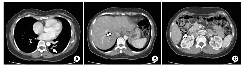
Figure 5 A follow up chest CT revealed decreased irregular necrotic mass (white arrows) in the lung (A), liver (B), and pancreas head (C). CT: computed tomography.
Reference
-
1. Golden MP, Vikram HR. Extrapulmonary tuberculosis: an overview. Am Fam Physician. 2005. 72:1761–1768.2. Yoon HJ, Song YG, Park WI, Choi JP, Chang KH, Kim JM. Clinical manifestations and diagnosis of extrapulmonary tuberculosis. Yonsei Med J. 2004. 45:453–461.3. Harisinghani MG, McLoud TC, Shepard JA, Ko JP, Shroff MM, Mueller PR. Tuberculosis from head to toe. Radiographics. 2000. 20:449–470.4. Fan ZM, Zeng QY, Huo JW, Bai L, Liu ZS, Luo LF, et al. Macronodular multi-organs tuberculoma: CT and MR appearances. J Gastroenterol. 1998. 33:285–288.5. Yu RS, Zhang SZ, Wu JJ, Li RF. Imaging diagnosis of 12 patients with hepatic tuberculosis. World J Gastroenterol. 2004. 10:1639–1642.6. Hwang SW, Kim YJ, Cho EJ, Choi JK, Kim SH, Yoon JH, et al. Clinical features of hepatic tuberculosis in biopsy-proven cases. Korean J Hepatol. 2009. 15:159–167.7. Kawamori Y, Matsui O, Kitagawa K, Kadoya M, Takashima T, Yamahana T. Macronodular tuberculoma of the liver: CT and MR findings. AJR Am J Roentgenol. 1992. 158:311–313.8. Gelb AF, Leffler C, Brewin A, Mascatello V, Lyons HA. Miliary tuberculosis. Am Rev Respir Dis. 1973. 108:1327–1333.9. Global tuberculosis control report 2010. World Health Organization (WHO). c2010. cited 2011 Jan 10. Geneva, Switzerland: WHO;Available from: http://whqlibdoc.who.int/publications/2010/9789241564069_eng.pdf.10. Sharma MP, Bhatia V. Abdominal tuberculosis. Indian J Med Res. 2004. 120:305–315.11. Kok KY, Yapp SK. Isolated hepatic tuberculosis: report of five cases and review of the literature. J Hepatobiliary Pancreat Surg. 1999. 6:195–198.12. Cho SB. Pancreatic tuberculosis presenting with pancreatic cystic tumor: a case report and review of the literature. Korean J Gastroenterol. 2009. 53:324–328.13. Ariyürek MO, Karçaaltincaba M, Demirkazik FB, Akay H, Gedikoglu G, Emri S. Bilateral multiple pulmonary tuberculous nodules mimicking metastatic disease. Eur J Radiol. 2002. 44:33–36.14. Song SH, Hahn HS, Kyung SY, Hwang JK, An CH, Lim YH, et al. A study of clinical investigations of pulmonary tuberculoma. Tuberc Respir Dis. 2002. 52:330–337.
- Full Text Links
-
- Actions
-
Cited
- CITED
-
- Close
- Share
-
- Similar articles
-
- A Case of Tuberculosis-related Retinal Vasculitis
- Cushing Syndrome Caused by ACTH-independent Macronodular Adrenal Hyperplasia
- Tuberculosis Verrucosa Cutis in a Patient with Pulmonary Tuberculosis
- A Case of Bilateral Macronodular Adrenocortical Hyperplasia
- A Study on the Drug Susceptibility Test of Multi-Drug Resistant Tuberculosis Patients
