Diagnosis and treatment of teeth with primary endodontic lesions mimicking periodontal disease: three cases with long-term follow ups
- Affiliations
-
- 1Department of Oral and Maxillofacial Surgery, Korea University Ansan Medical Hospital, Ansan, Korea.
- 2Division of Biomaterials and Bioengineering, Department of Preventive and Restorative Dental Sciences, University of California, San Francisco, CA, USA.
- 3Department of Conservative Dentistry, Yonsei University College of Dentistry, Seoul, Korea. shujungshin@yahoo.com
Abstract
- A tooth with primary endodontic disease that demonstrates a periodontal defect might be extracted because of misdiagnosis as severe periodontal disease or a vertical root fracture. The aim of this case report was to demonstrate the long-term survival of endodontically treated teeth, which had been initially considered unsavable. With meticulous evaluation including the patient's dental history, clinical and radiographic examinations, teeth with primary endodontic lesions could be differentiated and saved after proper root canal treatment. Pain history, vitality test, and radiographic examinations, as well as a general periodontal condition check with periodontal probing on an affected tooth, might be the key methods to differentiate endodontic pathosis from that of periodontal disease.
MeSH Terms
Figure
-
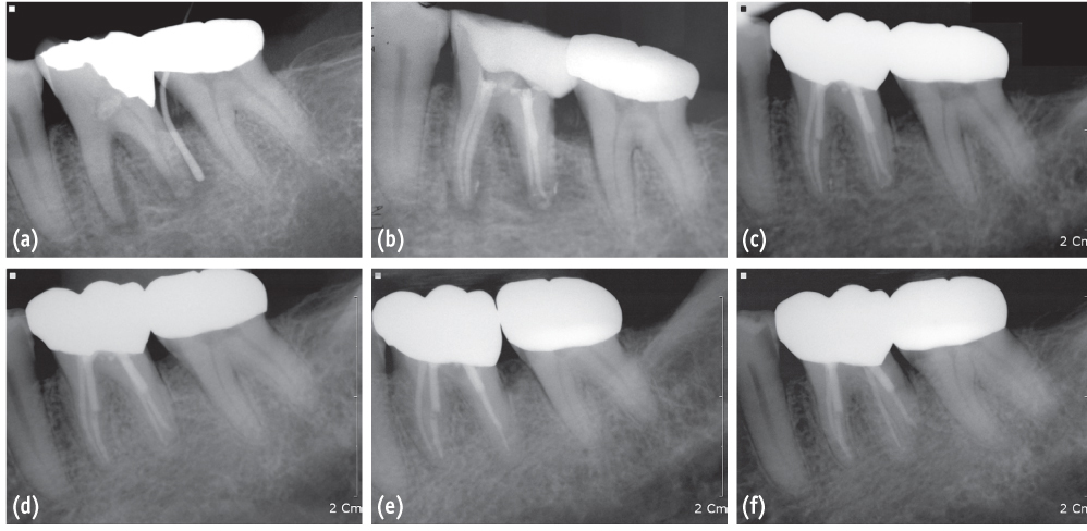
Figure 1 A series of periapical radiographs of the mandibular left first molar of case 1. (a) A preoperative radiograph showed radiolucency around both the mesial and distal roots and a gutta percha cone traced to the apex of distal root; (b) A postoperative radiograph after obturation of 4 canals; (c) A periapical radiograph at a 1-year routine check-up; (d) A periapical radiograph at a 3-year routine check-up; (e) and (f) Periapical radiographs at an 8-year routine check-up.
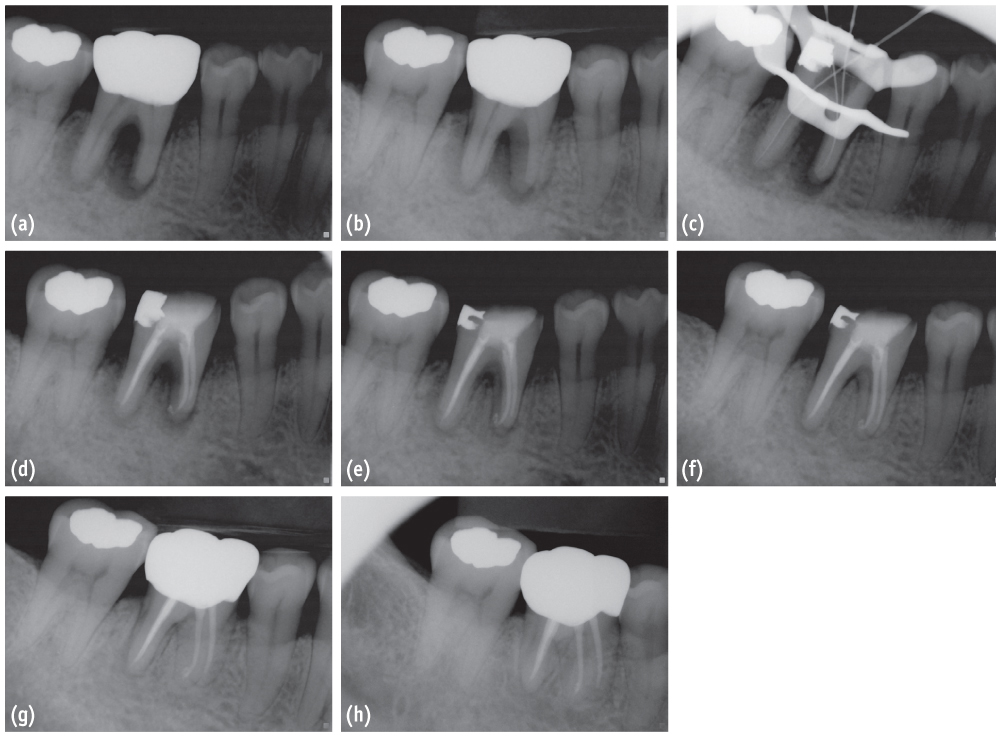
Figure 2 Periapical radiographs of case 2. (a) A preoperative periapical radiograph showed a J-shaped radiolucency on the mesial root of mandibular right first molar; (b) A preoperative radiograph with a different horizontal angle. The distal root also had a J-shaped lesion, which could be suspected of being a vertical root fracture; (c) A periapical radiograph for the working length determination; (d) A postoperative radiograph after canal filling; (e) A periapical radiograph at a 3-month routine check-up; (f) A periapical radiograph at a 6-month routine check-up; (g) A periapical radiograph at a 1-year routine check-up; (h) A periapical radiograph at a 2-year routine check-up.
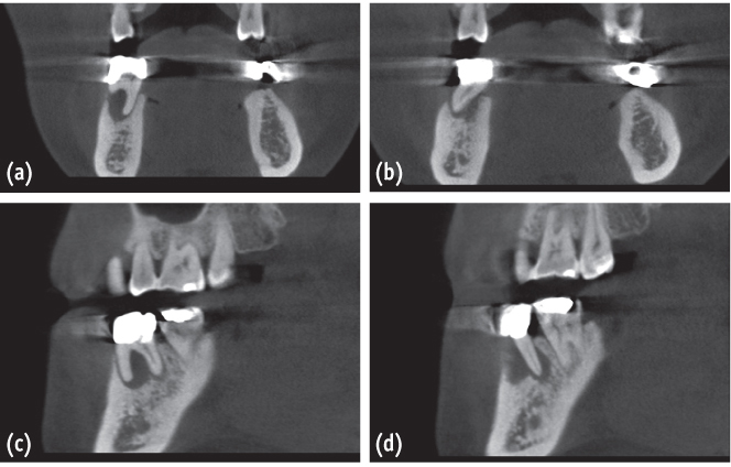
Figure 3 Preoperative views of #46 of case 2 using cone-beam computed tomography (CBCT). (a) A coronal view of the mesial root and the surrounding bone of #46; (b) A coronal view of the distal root and the surrounding bone of #46; (c) A sagittal view of the mesial root and the surrounding bone of #46; (d) A sagittal view of the distal root and the surrounding bone of #46.
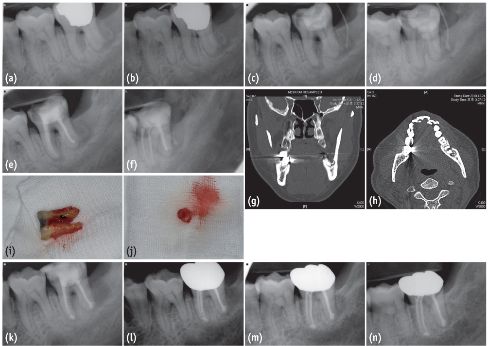
Figure 4 A series of periapical radiographs, computed tomographic (CT) views, and clinical photos of the mandibular left second molar of case 3. (a) A preoperative periapical radiograph of #37; (b) A preoperative radiograph of #37 with a gutta percha cone traced to the furcation area; (c) and (d) Periapical radiographs obtained at a second visit of the endodontic treatment of #37. A sinus tract formed and was traced to the distal surface of the tooth; (e) and (f) Postoperative radiographs of #37 after canal filling; (g) and (h) CT views from sagittal and axial planes demonstrated the tooth fragment of #38; (i) An extracted second molar for intentional replantation. Calculus deposition was noticed on the furcation of the distal root; (j) A tooth fragment of #38 was removed through an extraction socket of #37; (k) A postoperative radiograph after intentional replantation of #37; (l) A periapical radiograph at a 1-year routine check-up; (m) and (n) A periapical radiograph at a 2-year routine check-up.
Reference
-
1. Simon JH, Glick DH, Frank AL. The relationship of endodontic-periodontic lesions. J Endod. 2013; 39:e41–e46.
Article2. Harrington GW. The perio-endo question: differential diagnosis. Dent Clin North Am. 1979; 23:673–690.3. Chen SY, Wang HL, Glickman GN. The influence of endodontic treatment upon periodontal wound healing. J Clin Periodontol. 1997; 24:449–456.
Article4. Bergenholtz G. Interactions between pulpal and periodontal disease conditions: introduction. Endod Topics. 2006; 13:1–2.
Article5. Rotstein I, Simon JH. The endo-perio lesion: a critical appraisal of the disease condition. Endod Topics. 2006; 13:34–56.
Article6. Jansson LE, Ehnevid H. The influence of endodontic infection on periodontal status in mandibular molars. J Periodontol. 1998; 69:1392–1396.
Article7. Guldener PH. The relationship between periodontal and pulpal disease. Int Endod J. 1985; 18:41–54.
Article8. Harrington GW, Steiner DR, Ammons WF. The periodontal-endodontic controversy. Periodontol 2000. 2002; 30:123–130.
Article9. Sigurdsson A. Pulpal diagnosis. Endod Topics. 2003; 5:12–25.
Article10. Tamse A. Vertical root fractures in endodontically treated teeth: diagnostic signs and clinical management. Endod Topics. 2006; 13:84–94.
Article11. Pitts DL, Natkin E. Diagnosis and treatment of vertical root fractures. J Endod. 1983; 9:338–346.
Article12. Keesee SM, Baty DL, Cameron SM, Lefler TB, Morris WJ. A technique for achieving prerestorative minor tooth movement with orthodontic separators. J Prosthet Dent. 2002; 88:544–547.
Article13. Shin H, Roh BD, Shin YS, Lee CY. Pre-prosthetic minor tooth movement with elastic separating ring & provisional restoration modification: case report. Restor Dent Endod. 2012; 37:114–118.
Article
- Full Text Links
-
- Actions
-
Cited
- CITED
-
- Close
- Share
-
- Similar articles
-
- Endodontic treatment enhances the regenerative potential of teeth with advanced periodontal disease with secondary endodontic involvement
- Management of failed periodontal surgical intervention for a furcal lesion with a nonsurgical endodontic approach
- Recognition and management of palatogingival groove for tooth survival: a literature review
- Endodontic flare-ups incidence and related factors
- Guided tissue regeneration therapy after root canal therapy for long standing periodontal-endodontic combined lesion in the mandibular anterior area: case report
