Restor Dent Endod.
2012 Nov;37(4):201-206.
A survey of experience-based preference of Nickel-Titanium rotary files and incidence of fracture among general dentists
- Affiliations
-
- 1Department of Conservative Dentistry, Seoul National University School of Dentistry and Dental Research Institute, Seoul, Korea.
- 2Department of Conservative Dentistry, Yonsei University College of Dentistry, Seoul, Korea.
- 3Department of Statistics, Pusan National University, Busan, Korea.
- 4Department of Conservative Dentistry, Pusan National University School of Dentistry, Yangsan, Korea. golddent@pusan.ac.kr
Abstract
OBJECTIVES
The purpose was to investigate the preference and usage technique of NiTi rotary instruments and to retrieve data on the frequency of re-use and the estimated incidence of file separation in the clinical practice among general dentists.
MATERIALS AND METHODS
A survey was disseminated via e-mail and on-site to 673 general dentists. The correlation between the operator's experience or preferred technique and frequency of re-use or incidence of file fracture was assessed.
RESULTS
A total of 348 dentists (51.7%) responded. The most frequently used NiTi instruments was ProFile (39.8%) followed by ProTaper. The most preferred preparation technique was crown-down (44.6%). 54.3% of the respondents re-used NiTi files more than 10 times. There was a significant correlation between experience with NiTi files and the number of reuses (p = 0.0025). 54.6% of the respondents estimated experiencing file separation less than 5 times per year. The frequency of separation was significantly correlated with the instrumentation technique (p = 0.0003).
CONCLUSIONS
A large number of general dentists in Korea prefer to re-use NiTi rotary files. As their experience with NiTi files increased, the number of re-uses increased, while the frequency of breakage decreased. Operators who adopt the hybrid technique showed less tendency of separation even with the increased number of re-use.
Keyword
Figure
-
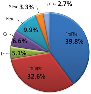
Figure 1 Preference of NiTi rotary instruments.
Reference
-
1. Walia HM, Brantley WA, Gerstein H. An initial investigation of the bending and torsional properties of Nitinol root canal files. J Endod. 1988. 14:346–351.
Article2. Schäfer E, Schulz-Bongert U, Tulus G. Comparison of hand stainless steel and nickel titanium rotary instrumentation: a clinical study. J Endod. 2004. 30:432–435.
Article3. da Silva FM, Kobayashi C, Suda H. Analysis of forces developed during mechanical preparation of extracted teeth using RaCe rotary instruments and ProFiles. Int Endod J. 2005. 38:17–21.
Article4. Shen Y, Cheung GS, Bian Z, Peng B. Comparison of defects in ProFile and ProTaper systems after clinical use. J Endod. 2006. 32:61–65.
Article5. Parashos P, Gordon I, Messer HH. Factors influencing defects of rotary nickel-titanium endodontic instruments after clinical use. J Endod. 2004. 30:722–725.
Article6. Xu X, Eng M, Zheng Y, Eng D. Comparative study of torsional and bending properties for six models of nickel-titanium root canal instruments with different cross-sections. J Endod. 2006. 32:372–375.
Article7. Kim HC, Kim HJ, Lee CJ, Kim BM, Park JK, Versluis A. Mechanical response of nickel-titanium instruments with different cross-sectional designs during shaping of simulated curved canals. Int Endod J. 2009. 42:593–602.
Article8. Turpin YL, Chagneau F, Bartier , Cathelineau G, Vulcain JM. Impact of torsional and bending inertia on root canal instruments. J Endod. 2001. 27:333–336.
Article9. Kim HC, Yum J, Hur B, Cheung GS. Cyclic fatigue and fracture characteristics of ground and twisted nickel-titanium rotary files. J Endod. 2010. 36:147–152.
Article10. Morgan LF, Montgomery S. An evaluation of the crown-down pressureless technique. J Endod. 1984. 10:491–498.
Article11. Walsch H. The hybrid concept of nickel-titanium rotary instrumentation. Dent Clin North Am. 2004. 48:183–202.
Article12. Schäfer E, Erler M, Dammaschke T. Comparative study on the shaping ability and cleaning efficiency of rotary Mtwo instruments. Part 2. Cleaning effectiveness and shaping ability in severely curved root canals of extracted teeth. Int Endod J. 2006. 39:203–212.
Article13. Grande NM, Plotino G, Pecci R, Bedini R, Malagnino VA, Somma F. Cyclic fatigue resistance and three-dimensional analysis of instruments from two nickel-titanium rotary systems. Int Endod J. 2006. 39:755–763.
Article14. Roland DD, Andelin WE, Browning DF, Hsu GH, Torabinejad M. The effect of preflaring on the rates of separation for 0.04 taper nickel titanium rotary instruments. J Endod. 2002. 28:543–545.
Article15. Lee M, Winkler J, Hartwell G, Stewart J, Caine R. Current trends in endodontic practice: emergency treatments and technological armamentarium. J Endod. 2009. 35:35–39.
Article16. Bird DC, Chambers D, Peters OA. Usage parameters of nickel-titanium rotary instruments: a survey of endodontists in the United States. J Endod. 2009. 35:1193–1197.
Article17. Madarati AA, Watts DC, Qualtrough AJ. Opinions and attitudes of endodontists and general dental practitioners in the UK towards the intracanal fracture of endodontic instruments: part 1. Int Endod J. 2008. 41:693–701.
Article18. Arens FC, Hoen MM, Steiman HR, Dietz GC Jr. Evaluation of single-use rotary nickel-titanium instruments. J Endod. 2003. 29:664–666.
Article19. Iqbal MK, Kohli MR, Kim JS. A retrospective clinical study of incidence of root canal instrument separation in an endodontics graduate program: a PennEndo database study. J Endod. 2006. 32:1048–1052.
Article20. Wolcott S, Wolcott J, Ishley D, Kennedy W, Johnson S, Minnich S, Meyers J. Separation incidence of protaper rotary instruments: a large cohort clinical evaluation. J Endod. 2006. 32:1139–1141.
Article21. Wu J, Lei G, Yan M, Yu Y, Yu J, Zhang G. Instrument separation analysis of multi-used ProTaper Universal rotary system during root canal therapy. J Endod. 2011. 37:758–763.
Article22. Parashos P, Messer HH. Questionnaire survey on the use of rotary nickel-titanium endodontic instruments by Australian dentists. Int Endod J. 2004. 37:249–259.
Article23. Lydeard S. The questionnaire as a research tool. Fam Pract. 1991. 8:84–91.
Article24. Hovland EJ, Romberg E, Moreland EF. Nonresponse bias to mail survey questionnaires within a professional population. J Dent Educ. 1980. 44:270–274.
Article25. Barbakow F, Lutz F. The 'Lightspeed' preparation technique evaluated by Swiss clinicians after attending continuing education courses. Int Endod J. 1997. 30:46–50.
Article26. Ounsi HF, Salameh Z, Al-Shalan T, Ferrari M, Grandini S, Pashley DH, Tay FR. Effect of clinical use on the cyclic fatigue resistance of ProTaper nickel-titanium rotary instruments. J Endod. 2007. 33:737–741.
Article27. Yared GM, Bou Dagher FE, Machtou P. Cyclic fatigue of ProFile rotary instruments after clinical use. Int Endod J. 2000. 33:204–207.
Article28. Sattapan B, Nervo GJ, Palamara JE, Messer HH. Defects in rotary nickel-titanium files after clinical use. J Endod. 2000. 26:161–165.
Article29. Moore J, Fitz-Walter P, Parashos P. A micro-computed tomographic evaluation of apical root canal preparation using three instrumentation techniques. Int Endod J. 2009. 42:1057–1064.
Article30. Setzer FC, Kwon TK, Karabucak B. Comparison of apical transportation between two rotary file systems and two hybrid rotary instrumentation sequences. J Endod. 2010. 36:1226–1229.
Article31. Kim JW, Park JK, Hur B, Kim HC. Comparison of shaping ability using various Nickel-Titanium rotary files and hybrid technique. J Korean Acad Conserv Dent. 2007. 32:530–541.
Article32. Sonntag D, Ott M, Kook K, Stachniss V. Root canal preparation with the NiTi systems K3, Mtwo and ProTaper. Aust Endod J. 2007. 33:73–81.
Article
- Full Text Links
-
- Actions
-
Cited
- CITED
-
- Close
- Share
-
- Similar articles
-
- Shaping ability of Ni-Ti rotary files in combination with GT rotary Ni-Ti file
- Influence of root canal curvature on the screw-in effect of nickel-titanium rotary files in simulated resin root canal
- Mechanical and geometric features of endodontic instruments and its clinical effect
- Preference of undergraduate students after first experience on nickel-titanium endodontic instruments
- Effect of various canal preparation techniques using rotary nickel-titanium files on the maintenance of canal curvature
