Korean J Urol.
2013 Jul;54(7):482-485.
Bilateral Single Ectopic Ureters Draining Into a Grossly Dilated Vagina in an Adolescent Female
- Affiliations
-
- 1Department of Urology, Calcutta National Medical College and Hospital, Kolkata, India. gk_amc@yahoo.co.in
- 2Department of Urology, Kolkata, R.G. Kar Medical College and Hospital, Kolkata, India.
Abstract
- A 16-year-old female presented with dribbling of urine along with voluntary voiding since birth. Renal imaging revealed hydroureteronephrosis on the right side; the uterus and ovary were normal. A radionuclide scan showed a left nonfunctional kidney. On cystovaginoscopy, the urethra was shown to be normal and the urinary bladder was tubular with small capacity and an absent trigone. Although the vagina was capacious, no ureteric orifices were found. Computed tomography corroborated the diagnosis of bilateral, single ectopic ureters draining into a grossly dilated vagina. This case is unique because it is a bilateral single-system ureteral ectopia in a completely differentiated female genital tract that presented late in adolescence. To the best of our knowledge, this is the second such ureteral abnormality reported in the literature so far. The patient underwent ileocystoplasty with right ureteric reimplantation and nephroureterectomy for the left nonfunctional kidney, which histopathology showed to be tuberculosis. The patient is continent with cystometric capacity of more than 300 mL.
Keyword
MeSH Terms
Figure
-
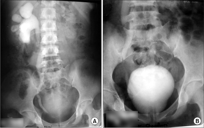
FIG. 1 (A) Intravenous urogram (IVU) showing right hydroureteronephrosis with contrast filling the capacious vagina. (B) IVU: full apparent urinary bladder (vagina).

FIG. 2 (A) Contrast computed tomography (CT) showing grossly dilated vagina (V) with Foley's bulb (F) in situ, anteriorly displaced uterus (U), and rectum (R) posteriorly. (B) Subsequent image to Fig 2A. (C) CT image showing relation of distal vagina (V) with small urinary bladder (UB) and anorectum (AR). UT, uterus.

FIG. 3 Intraoperative picture.

FIG. 4 (A) Micturating cystourethrogram (MCU) showing reconstructed bladder postileocystoplasty. (B) MCU: postvoid film with adequate emptying.
Reference
-
1. Chatterjee US, Chatterjee SK. Novel bladder augmentation in a bilateral single system vaginal ectopia. Afr J Paediatr Surg. 2011. 8:109–111.2. Sheldon CA, Welch TR. Total anatomic urinary tract replacement and renal transplantation: a surgical strategy to correct severe genitourinary anomalies. J Pediatr Surg. 1998. 33:635–638.3. Singh BP, Pathak HR, Andankar MG. Bilateral single-system ectopic ureters opening into vaginalized urogenital sinus. Indian J Urol. 2010. 26:126–128.4. Pantuck AJ, Barone JG, Rosenfeld DL, Fleisher MH. Occult bilateral ectopic vaginal ureters causing urinary incontinence: diagnosis by computed tomography. Abdom Imaging. 1996. 21:78–80.5. Wakhlu A, Dalela D, Tandon RK, Chandra H, Wakhlu AK. The single ectopic ureter. Br J Urol. 1998. 82:246–251.6. Podesta E, Scarsi PL, Di Rovasenda E, Ferretti S, Magillo P, Dodero P. Vesical continence in bilateral ectopic single ureters. J Urol. 2001. 165(6 Pt 2):2363–2365.
- Full Text Links
-
- Actions
-
Cited
- CITED
-
- Close
- Share
-
- Similar articles
-
- Single Vaginal Ectopic Ureter: A Case Report
- Ectopic Ureteral Orifice Associated with Ipsilateral Renal Dysplasia
- Two Cases of Single Ectopic Ureter Associated with Ectopic Renal Dysplasia
- Management of Duplicated Ureters with the Ectopic Ureter Opening into the Vaginal Vestibule by Ureter Opening into the Vaginal Vestibule by Pyeloureterostomy: A Case Report
- A Case of Giant Hydronephrosis with Ectopic Ureterocele in Bilateral Duplicated Ureters
