Korean J Urol.
2013 Oct;54(10):660-665.
Experience of Ultrasonography-Guided Percutaneous Core Biopsy for Renal Masses
- Affiliations
-
- 1Department of Urology, Soonchunhyang University Cheonan Hospital, Cheonan, Korea. ysurol@schmc.ac.kr
- 2Department of Urology, Soonchunhyang University Seoul Hospital, Seoul, Korea.
Abstract
- PURPOSE
We evaluated the safety and accuracy of ultrasonography-guided percutaneous core biopsy collection in patients with renal masses.
MATERIALS AND METHODS
From June 2008 to August 2012, 30 percutaneous core biopsies of renal masses were performed. The biopsies obtained were small tumors (<4 cm) with ambiguous radiologic findings or that met classic renal biopsy indications. The biopsy results were compared with the final pathological results after definitive surgical treatment. Ultrasonography was performed on the day after biopsy collection to rule out any complications.
RESULTS
The mean age of the patients was 57.7 years, and the mean tumor size was 3.39 cm. Twelve of the lesions were in the left kidney, and 18 were in the right kidney. All but one core biopsy contained sufficient material for histopathological analysis. The biopsy results showed 17 renal cell carcinomas (56.7%), 3 angiomyolipomas (10.0%), 2 oncocytomas (6.7%), 1 adenocarcinoma (3.3%), and 7 benign lesions (23.3%). A total of 18 cases underwent surgery, and the pathological results confirmed the initial biopsy diagnosis for 17 of 18 cases (94.4%). The one (5.9%) inaccurate biopsy result was found to be a urothelial carcinoma of the kidney. No needle tract seeding was found in the pathological specimens or on follow-up imaging. A small perinephric hematoma (1-2 cm) was seen in 5 cases (16.7%), but all patients remained hemodynamically stable.
CONCLUSIONS
Ultrasonography-guided renal biopsy is a safe, effective, and accurate method for evaluating small renal masses. This procedure may help in selecting treatment modalities for small renal masses.
Keyword
MeSH Terms
Figure
-
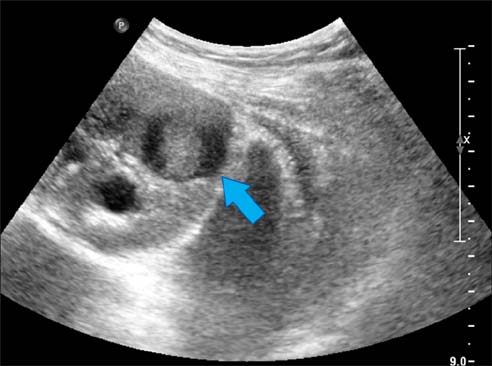
FIG. 1 Targeting the lesion (arrow indicated the mass).
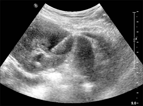
FIG. 2 At the moment of puncture the lesion.

FIG. 3 The flow chart of percutaneous renal biopsy patients. RCC, renal cell carcinoma; AML, angiomyolipoma.

FIG. 4 Small perirenal hematoma formation after biopsy.
Reference
-
1. Smith SJ, Bosniak MA, Megibow AJ, Hulnick DH, Horii SC, Raghavendra BN. Renal cell carcinoma: earlier discovery and increased detection. Radiology. 1989; 170(3 Pt 1):699–703.2. Frank I, Blute ML, Cheville JC, Lohse CM, Weaver AL, Zincke H. Solid renal tumors: an analysis of pathological features related to tumor size. J Urol. 2003; 170(6 Pt 1):2217–2220.3. Dechet CB, Zincke H, Sebo TJ, King BF, LeRoy AJ, Farrow GM, et al. Prospective analysis of computerized tomography and needle biopsy with permanent sectioning to determine the nature of solid renal masses in adults. J Urol. 2003; 169:71–74.4. Richter F, Kasabian NG, Irwin RJ Jr, Watson RA, Lang EK. Accuracy of diagnosis by guided biopsy of renal mass lesions classified indeterminate by imaging studies. Urology. 2000; 55:348–352.5. Lane BR, Samplaski MK, Herts BR, Zhou M, Novick AC, Campbell SC. Renal mass biopsy: a renaissance? J Urol. 2008; 179:20–27.6. Campbell SC, Lane BR. Malignant renal tumors. In : Wein AJ, Kavoussi LR, Novick AC, Partin AW, Peters CA, editors. Campbell's urology. 10th ed. Philadelphia: Saunders;2012. p. 1413–1474.7. Israel GM, Bosniak MA. How I do it: evaluating renal masses. Radiology. 2005; 236:441–450.8. Volpe A, Panzarella T, Rendon RA, Haider MA, Kondylis FI, Jewett MA. The natural history of incidentally detected small renal masses. Cancer. 2004; 100:738–745.9. Chawla SN, Crispen PL, Hanlon AL, Greenberg RE, Chen DY, Uzzo RG. The natural history of observed enhancing renal masses: meta-analysis and review of the world literature. J Urol. 2006; 175:425–431.10. Poisson JF, Mejean A, Hupertan V, Chretien Y, Dufour B, Thiounn N. Kidney tumours: single-centre study of 810 patients. Changing features over a period of 15 years. Prog Urol. 2005; 15:1056–1061.11. Wood BJ, Khan MA, McGovern F, Harisinghani M, Hahn PF, Mueller PR. Imaging guided biopsy of renal masses: indications, accuracy and impact on clinical management. J Urol. 1999; 161:1470–1474.12. Silverman SG, Gan YU, Mortele KJ, Tuncali K, Cibas ES. Renal masses in the adult patient: the role of percutaneous biopsy. Radiology. 2006; 240:6–22.13. Reichelt O, Gajda M, Chyhrai A, Wunderlich H, Junker K, Schubert J. Ultrasound-guided biopsy of homogenous solid renal masses. Eur Urol. 2007; 52:1421–1426.14. Lebret T, Poulain JE, Molinie V, Herve JM, Denoux Y, Guth A, et al. Percutaneous core biopsy for renal masses: indications, accuracy and results. J Urol. 2007; 178(4 Pt 1):1184–1188.15. Ralls PW, Barakos JA, Kaptein EM, Friedman PE, Fouladian G, Boswell WD, et al. Renal biopsy-related hemorrhage: frequency and comparison of CT and sonography. J Comput Assist Tomogr. 1987; 11:1031–1034.16. Neuzillet Y, Lechevallier E, Andre M, Daniel L, Coulange C. Accuracy and clinical role of fine needle percutaneous biopsy with computerized tomography guidance of small (less than 4.0 cm) renal masses. J Urol. 2004; 171:1802–1805.17. Smith EH. Complications of percutaneous abdominal fine-needle biopsy: review. Radiology. 1991; 178:253–258.
- Full Text Links
-
- Actions
-
Cited
- CITED
-
- Close
- Share
-
- Similar articles
-
- Imaging-Guided Biopsy, Percutaneous Ablation, and Active Surveillance for Small Renal Masses
- Benign core biopsy of probably benign breast lesions 2 cm or larger: correlation with excisional biopsy and long-term follow-up
- Management of Breast Masses Detected only by Ultrasonography
- Usefulness of US-Guided Automated Gun Biopsy of Neck Masses
- Comparison of Needle Size in Pediatric Renal Biopsy with Sono-Guided Percutaneous Automated Gun Technique
