Korean J Urol.
2013 Nov;54(11):772-777.
Predictive Value of Preoperative Unenhanced Computed Tomography During Ureteroscopic Lithotripsy: A Single Institute's Experience
- Affiliations
-
- 1Department of Urology, Kyung Hee University School of Medicine, Seoul, Korea. yookoohan@khu.ac.kr
- 2Department of Anesthesiology and Pain Medicine, Kyung Hee University School of Medicine, Seoul, Korea.
- 3Department of Surgery, Kyung Hee University School of Medicine, Seoul, Korea.
Abstract
- PURPOSE
Ureteroscopic stone removal is frequently used to remove ureteral stones. Mucosal edema and bleeding are the two most important obstacles to a successful operation. This study analyzed relationships between unenhanced computed tomography (UECT) findings and ureteroscopic findings to determine whether ureteroscopic results could be predicted preoperatively by using UECT imaging.
MATERIALS AND METHODS
From January 2009 to July 2011, 675 patients were diagnosed with ureteral stones through UECT. Among them, we retrospectively reviewed 92 cases of patients who underwent ureteroscopy (URS). We identified findings such as hydronephrosis, rim sign, periureteral fat stranding, and perinephric fat stranding on the UECT and then categorized these findings into four categories (none, mild, moderate, and severe) according to their severity. We also divided the URS findings of mucosal edema and bleeding into four categories (none, mild, moderate, and severe) and compared these findings with the UECT images.
RESULTS
A total of 92 study patients were included in this study: 59 were male and 33 were female patients. According to the location of the stone, 31 cases were classified as upper ureteral stones, 15 were midureteral stones, and 46 were lower ureteral stones. Hydronephrosis identified with UECT was correlated with the mucosal edema severity observed during URS (p=0.004). The rim signs identified with UECT were proportional to the grade of mucosal edema (p=0.010).
CONCLUSIONS
Hydronephrosis and rim signs observed during UECT can be used as a predictive factor for intraoperative mucosal edema in patients undergoing URS.
MeSH Terms
Figure
-
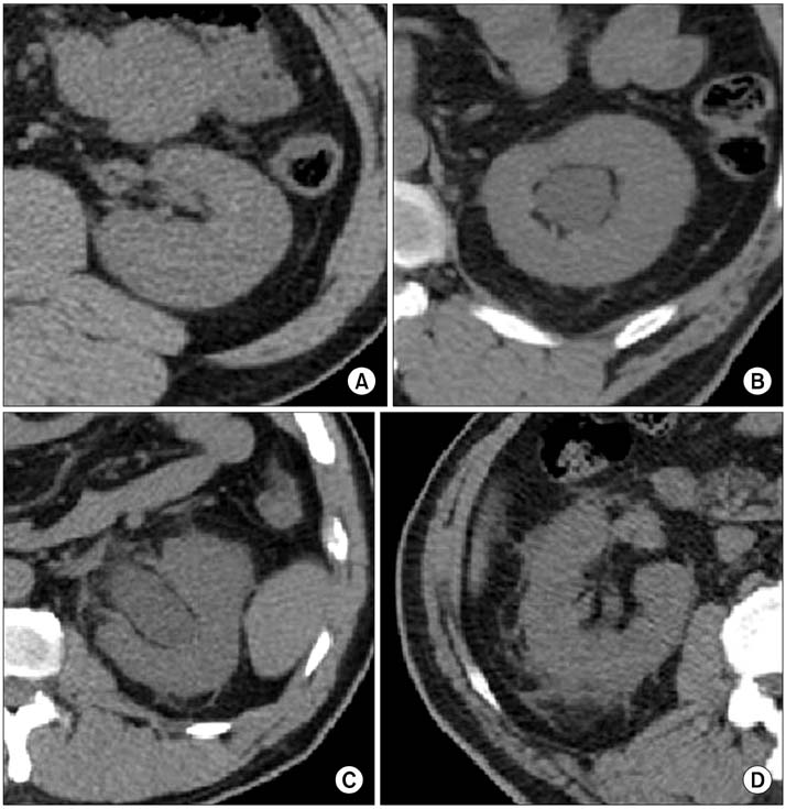
FIG. 1 Images taken from unenhenced computed tomography. Perinephric fat stranding was categorized according to its degree of perinephric fat stranding. A, none; B, mild; C, moderate; D, severe.
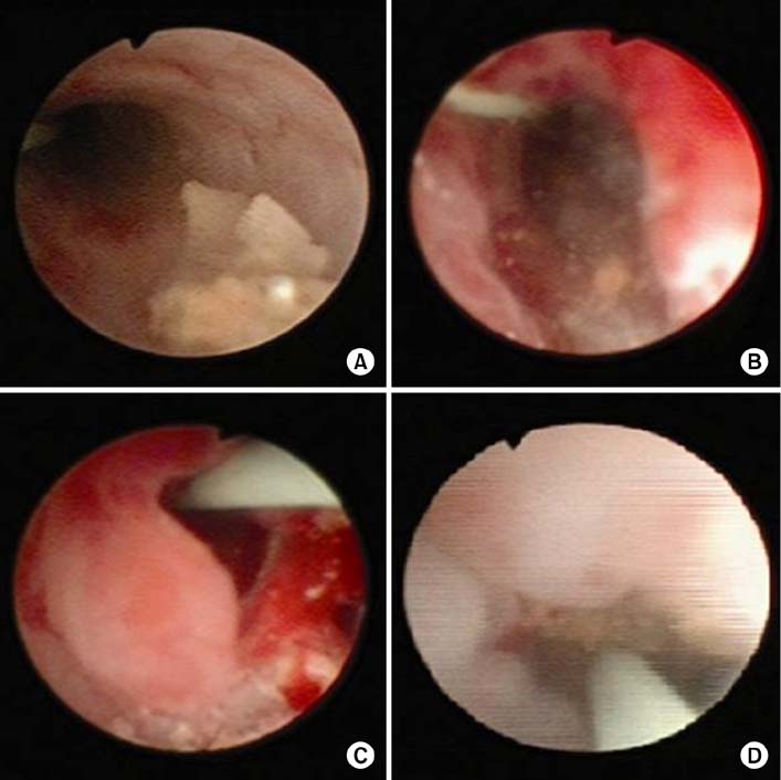
FIG. 2 Images taken during ureteroscopy. Mucosal edema was categorized according to its degree of mucosal edema. A, none; B, mild; C, moderate; D, severe.
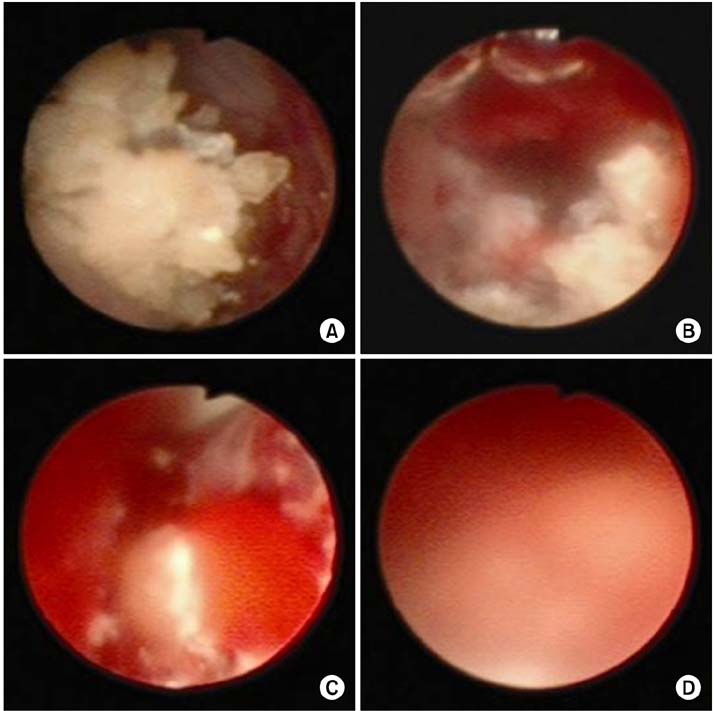
FIG. 3 Images taken during ureteroscopy. Intraluminal bleeding was categorized according to its degree of intraluminal bleeding. A, none; B, mild; C, moderate; D, severe.
Reference
-
1. Smith RC, Rosenfield AT, Choe KA, Essenmacher KR, Verga M, Glickman MG, et al. Acute flank pain: comparison of non-contrast-enhanced CT and intravenous urography. Radiology. 1995; 194:789–794.2. Dalrymple NC, Verga M, Anderson KR, Bove P, Covey AM, Rosenfield AT, et al. The value of unenhanced helical computerized tomography in the management of acute flank pain. J Urol. 1998; 159:735–740.3. Preminger GM, Tiselius HG, Assimos DG, Alken P, Buck C, Gallucci M, et al. 2007 guideline for the management of ureteral calculi. J Urol. 2007; 178:2418–2434.4. Abdelrahim AF, Abdelmaguid A, Abuzeid H, Amin M, Mousa el-S, Abdelrahim F. Rigid ureteroscopy for ureteral stones: factors associated with intraoperative adverse events. J Endourol. 2008; 22:277–280.5. Seitz C, Memarsadeghi M, Fajkovic H, Tanovic E. Secondary signs of non-enhanced CT prior to laser ureterolithotripsy: is treatment outcome predictable? J Endourol. 2008; 22:415–418.6. Anderson KR, Smith RC. CT for the evaluation of flank pain. J Endourol. 2001; 15:25–29.7. Alshamakhi AK, Barclay LC, Halkett G, Kukade G, Mundhada D, Uppoor RR, et al. CT evaluation of flank pain and suspected urolithiasis. Radiol Technol. 2009; 81:122–131.8. Smith RC, Verga M, Dalrymple N, McCarthy S, Rosenfield AT. Acute ureteral obstruction: value of secondary signs of helical unenhanced CT. AJR Am J Roentgenol. 1996; 167:1109–1113.9. Ege G, Akman H, Kuzucu K, Yildiz S. Acute ureterolithiasis: incidence of secondary signs on unenhanced helical CT and influence on patient management. Clin Radiol. 2003; 58:990–994.10. Smith RC, Dalrymple NC, Neitlich J. Noncontrast helical CT in the evaluation of acute flank pain. Abdom Imaging. 1998; 23:10–16.11. Fielding JR, Fox LA, Heller H, Seltzer SE, Tempany CM, Silverman SG, et al. Spiral CT in the evaluation of flank pain: overall accuracy and feature analysis. J Comput Assist Tomogr. 1997; 21:635–638.12. Herrera-Gonzalez G, Netsch C, Oberhagemann K, Bach T, Gross AJ. Effectiveness of single flexible ureteroscopy for multiple renal calculi. J Endourol. 2011; 25:431–435.13. Ghalayini IF, Al-Ghazo MA, Khader YS. Extracorporeal shockwave lithotripsy versus ureteroscopy for distal ureteric calculi: efficacy and patient satisfaction. Int Braz J Urol. 2006; 32:656–664.14. Strohmaier WL, Schubert G, Rosenkranz T, Weigl A. Comparison of extracorporeal shock wave lithotripsy and ureteroscopy in the treatment of ureteral calculi: a prospective study. Eur Urol. 1999; 36:376–379.15. el-Sherif AE. Endoscopic management of impacted stones in the intramural or meatal part of the ureter without performing meatotomy. Br J Urol. 1995; 76:394–396.16. Morgentaler A, Bridge SS, Dretler SP. Management of the impacted ureteral calculus. J Urol. 1990; 143:263–266.17. Lingeman JE, Shirrell WL, Newman DM, Mosbaugh PG, Steele RE, Woods JR. Management of upper ureteral calculi with extracorporeal shock wave lithotripsy. J Urol. 1987; 138:720–723.18. Dalrymple NC, Casford B, Raiken DP, Elsass KD, Pagan RA. Pearls and pitfalls in the diagnosis of ureterolithiasis with unenhanced helical CT. Radiographics. 2000; 20:439–447.
- Full Text Links
-
- Actions
-
Cited
- CITED
-
- Close
- Share
-
- Similar articles
-
- The Initial Experience with Transurethral Ureteroscopic Lithotripsy
- Clinical factors associated with postoperative hydronephrosis after ureteroscopic lithotripsy
- The Comparison of Ureteroscopic Lithotripsy Between Laser and Electrohydraulic Lithoriptor
- Clinical Experience with Ureteroscopic Management of Ureteral Calculi Including Electrohydraulic Lithotripsy
- Extracorporeal Shockwave Lithotripsy Versus Ureteroscopic Removal for Lower Ureteral Stones
