The Influence of Volume Effect in 2D-array Ion Chamber on the Measurement of IMRT Dose Distribution
- Affiliations
-
- 1Department of Radiation Oncology, Kyungpook National University Hospital, Daegu, Korea. jckim@knu.ac.kr
- 2Department of Radiation Oncology, Kyungpook National University School of Medicine, Daegu, Korea.
- 3Department of Physics, College of Science, Yeungnam University, Gyongsan, Korea.
Abstract
- We evaluated the influence of volume effect on the measurement of IMRT dose distribution by comparing a 2D-array ion chamber and other dosimeters. Matrix phantom which is a 2D-array ion chamber having volume effect was compared with beam image system and film for the measurement of dose distribution. Five intensity-modulated radiation therapy plans were created using five fields in thevirtual phantom. The measured dose distribution was compared with the calculated one by radiation treatment planning system and analysis program. We evaluated the conformity of dose distribution by calculating correlation coefficients and gamma values. The highest error rate of 1.3% was associated with matrix phantom in which volume effect in small field sizes was substantial.
Keyword
Figure
-
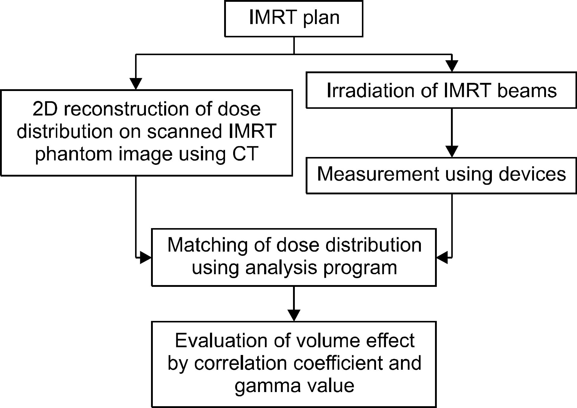
Fig. 1. Schematic overview of the various steps in the IMRT dose distribution measurement.
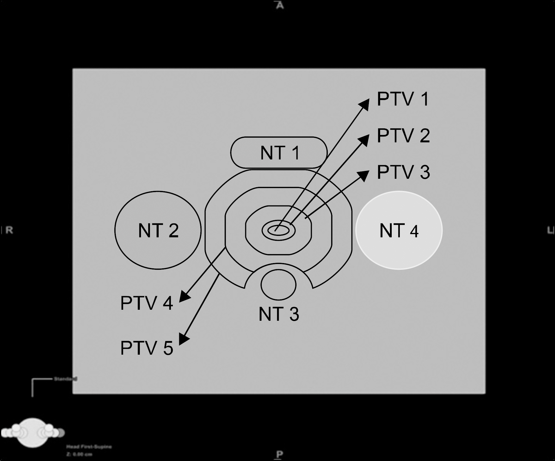
Fig. 2. Design of PTVs and normal tissues in the virtual phantom.
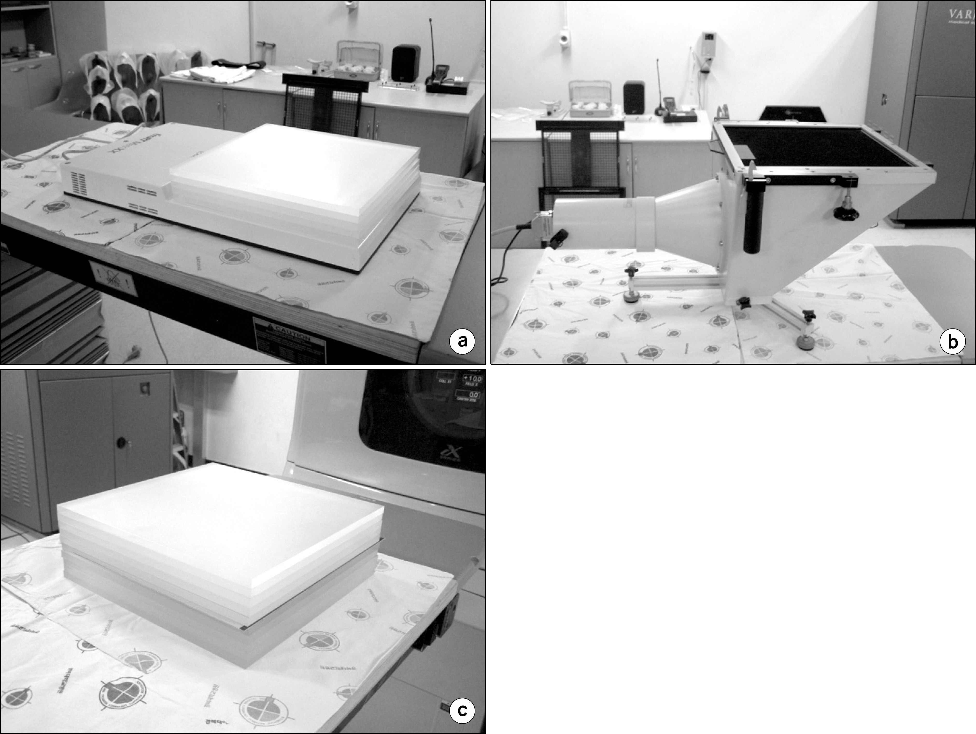
Fig. 3. Setup for measurement of dose distribution. (a) Matrix phantom. (b) BIS. (c) Film.
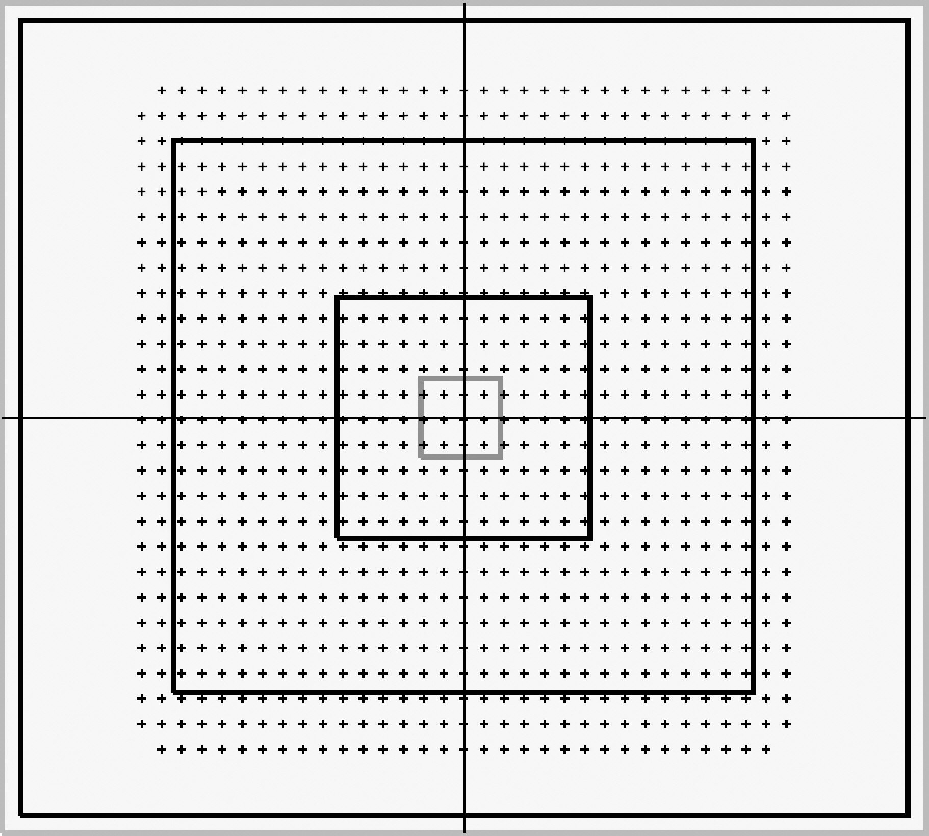
Fig. 4. Matrix phantom: 1,020 parallel plate chambers are on the matrix plate, spacing 0.762 mm internal.
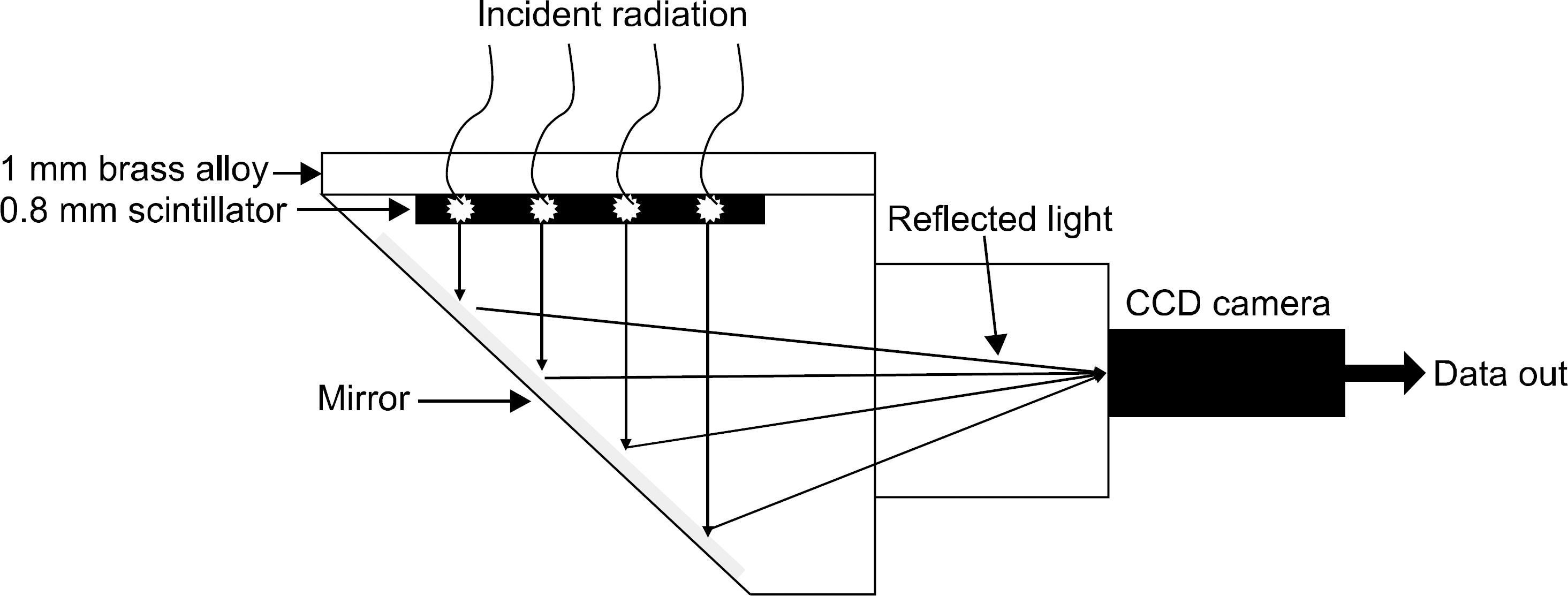
Fig. 5. Beam image system operated by the principle of scintillation consists of the following components: 1 mm brass alloy, 0.8 mm scintillator, mirror, ccd camera etc.

Fig. 6. The calculated correlation coefficient by analysis program with three different detectors in changeable side of square field.
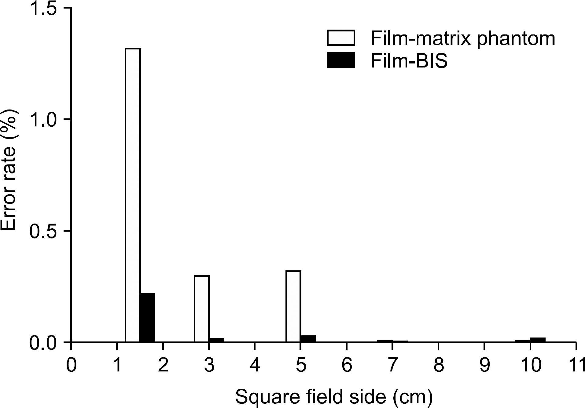
Fig. 7. Error rates with film-matrix phantom and film-BIS.
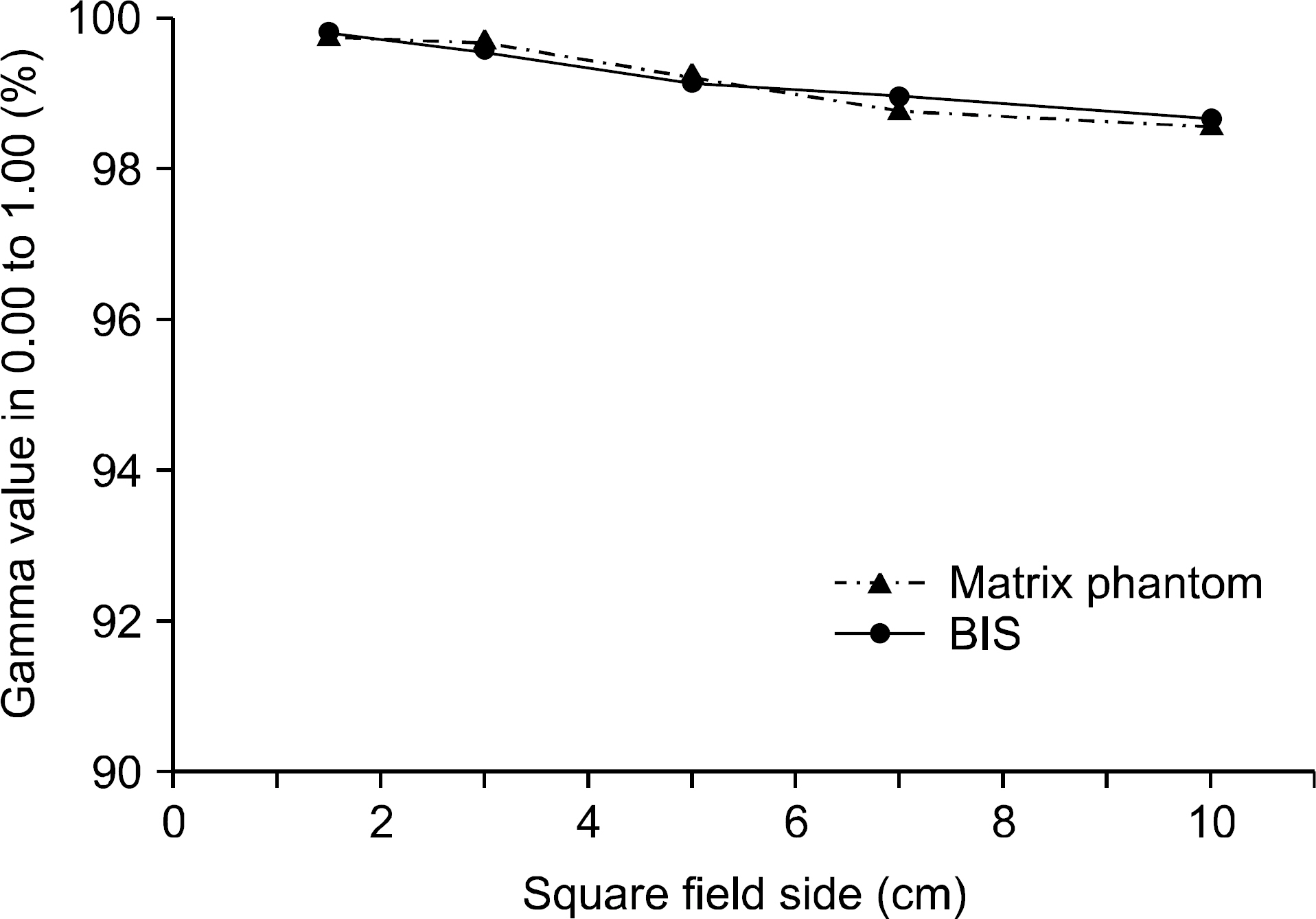
Fig. 8. The calculated gamma value by analysis program with Matrix phantom and BIS in changeable side of square field.
Reference
-
References
1. AAPM Report 82: Guidance document on delivery, treatment planning, and clinical implementation of IMRT. Report of the IMRT Subcommittee of the AAPM Radiation Therapy Committee. Med Phys. 30:2089–2115. 2003.2. Martens C, De Wagter C, De Neve W. The value of the PinPoint ion chamber for characterization of small field segments used in intensitymodulated radiotherapy. Phys Med Biol. 45:2519–2530. 2000.
Article3. Calcina CS, De Oliveira LN, Almeida CE, et al. Dosimetric parameters for small field sizes using Fricke xylenol gel, thermoluminescent and film dosimeters, and an ionization chamber. Phys Med Biol. 52:1431–1439. 2007.
Article4. Mack A, Scheib SG, Major J, et al. Precision dosimetry for narrow photon beams used in radiosurgery determination of gamma knife output factors. Med Phys. 29:2080–2089. 2002.5. Nizin PS. Eletronic equilibrium and primary dose in collimated photon beams: basic concepts and definitions. Med Phys. 26:1893–1900. 1993.6. Das IJ, Ding GX, Ahnesjö A. Small fields: Nonequilibrium radiation dosimetry. Med Phys. 35:206–215. 2008.
Article7. Laub WU, Wong T. The volume effect of detectors in the dosimetry of small field used in IMRT. Med Phys. 30:341–347. 2003.8. Sibata CH, Mota HC, Beddar AS, et al. Influence of detector size in photon beam profile measurements. Phys Med Biol. 36:621–631. 1991.
Article9. Alfonso R, Andreo P, Capote R, et al. A new formalism for reference dosimetry of small and nonstandard fields. Med Phys. 35:5179–5186. 2008.
Article10. Wong CJ, Ackerly T, He C, et al. Small field size dose-profile measurements using gel dosimeters, gafchromic films and micro-thermoluminescent dosimeters. Rad Meas. 44:249–256. 2009.
Article11. 고승영, 김성준: IMRT 및 IMRS에서 Field의 선량분포 확인시 SAD 변화에 따른 측정의 유용성 평가. 대한방사선치료학회지. 22:33–39. 2010.12. Lee JW, Hong SM, Kim YL, et al. Dosimetric characterization of ion chamber matrix for intensity modulated radiation therapy quality assurance. Korean J Med Phys. 17(3):131–135. 2006.13. Poppe B. Two-dimensional ionization chamber arrays for IMRT plan verification. Med phys. 33(4):1005–1015. 2006.
Article14. Vagovic P, Korytár D, Cecilia A, et al. High-resolution high-efficiency X-ray imaging system based on the in-line Bragg magnifier and the Medipix detector. Synchrotron Rad 20: Part1 (. 2013.15. Nikl M. Scintillation detectors for x-rays. Meas Sci Technol. 17:R37–R54. 2006.
Article16. I'mRT QA Hardware Manual: Image Device and Accessories. Scanditronix Wellhofer, p. .11 (. 2003.17. Omnipro I'mRT System Manual: Algorithms. Scanditronix Wellhofer, p. .203 (. 2008.18. Winiecki J, Morgas T, Majewska K, et al. The gamma evaluation method as a routine QA procedure of IMRT. Rep Pract Oncol Radiother. 14:162–168. 2009.
Article19. Ong CL, Cuijpers JP, Senan S, et al. Impact of the calculation resolution of AAA for small fields and RapidArc treatment plans. Med Phys. 38(8):4471–4479. 2011.
Article
- Full Text Links
-
- Actions
-
Cited
- CITED
-
- Close
- Share
-
- Similar articles
-
- Analysis on the Dosimetric Characteristics of Tangential Breast Intensity Modulated Radiotherapy
- Efficiency Study of 2D Diode Array Detector for IMRT Quality Assurance
- Dosimetric Analysis on the Effect of Target Motion in the Delivery of Conventional IMRT, RapidArc and Tomotherapy
- Development of Two-dimensional Prompt-gamma Measurement System for Verification of Proton Dose Distribution
- Dose Distribution of Intensity Modulated Radiation Therapy in Prostate Cancer
