Korean J Urol.
2009 Dec;50(12):1258-1261.
Aggressive Angiomyxoma as the Cause of Lower Urinary Tract Symptoms
- Affiliations
-
- 1Department of Urology, School of Medicine, Kyung Hee University, Seoul, Korea. sgchang@khu.ac.kr
- 2Department of Pathology, School of Medicine, Kyung Hee University, Seoul, Korea.
Abstract
- Aggressive angiomyxoma (AAM) is a rare, benign tumor. It usually involves the connective tissue of the perineal regions in women of reproductive age. In this report, we present a case of AAM in a 66-year-old female, which presented itself as a retrovesical tumor on pelvic magnetic resonance imaging and caused lower urinary tract symptoms. The tumor was resected en bloc and the patient's voiding symptoms disappeared.
Keyword
MeSH Terms
Figure
-

Fig. 1 (A, B) Transvaginal ultrasonography showing a huge tumor compressing the urinary bladder. (C) T1-weighted magnetic resonance image (MRI) showing a mass with low signal intensity posterior to the urinary bladder. (D) T2-weighted sagittal MRI showing a mass with intermediate signal intensity.
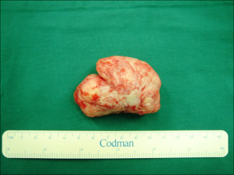
Fig. 2 The gross findings are a rather well-circumscribed gray-white mass with a soft, smooth and elastic surface.
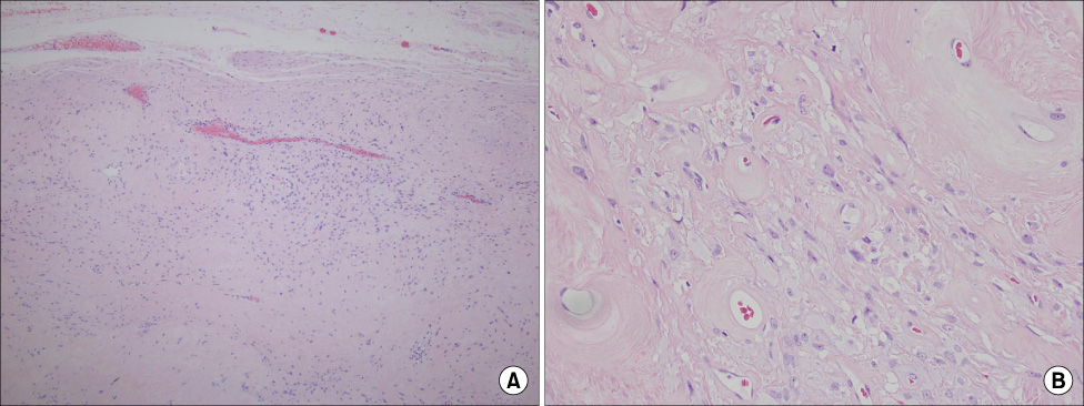
Fig. 3 Pathologic findings show dispersed spindle-shaped cells and thick-walled blood vessels with a myxoid stroma [(A) ×100, (B) ×400].
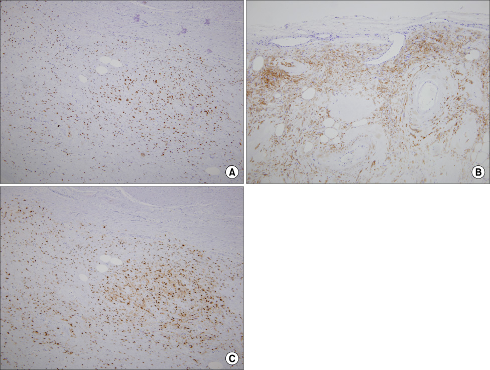
Fig. 4 Immunohistochemical analyses show positive results for (A) estrogen receptor, (B) desmin, and (C) progesterone receptor.
Reference
-
1. Fetsch JF, Laskin WB, Lefkowitz M, Kindblom LG, Meis-Kindblom JM. Aggressive angiomyxoma: a clinicopathologic study of 29 female patients. Cancer. 1996. 78:79–90.2. Hatano K, Tsujimoto Y, Ichimaru N, Miyagawa Y, Nonomura N, Okuyama A. Rare case of aggressive angiomyxoma presenting as a retrovesical tumor. Int J Urol. 2006. 13:1012–1014.3. Lee CJ, Yun GB, Kim HY, Kim WG, Kim SH, Park ED, et al. Aggressive angiomyxoma of the vagina after menopause. Korean J Gynecol Oncol Colposc. 2003. 14:167–171.4. Steeper TA, Rosai J. Aggressive angiomyxoma of the female pelvis and perineum. Report of nine cases of a distinctive type of gynecologic soft-tissue neoplasm. Am J Surg Pathol. 1983. 7:463–475.5. Kim TH, Kim YS, Myung SC, Shin HO, Lee TJ, Yoo SM, et al. An aggressive, rapidly increasing angiomyxoma of the scrotum. Korean J Urol. 2006. 47:553–555.6. Jeyadevan NN, Sohaib SA, Thomas JM, Jeyarajah A, Shepherd JH, Fisher C. Imaging features of aggressive angiomyxoma. Clin Radiol. 2003. 58:157–162.7. Smith HO, Worrell RV, Smith AY, Dorin MH, Rosenberg RD, Bartow SA. Aggressive angiomyxoma of the female pelvis and perineum: review of the literature. Gynecol Oncol. 1991. 42:79–85.8. Smirniotis V, Kondi-Pafiti A, Theodoraki K, Kostopanagiotou G, Liapis A, Kourias E. Aggressive angiomyxoma of the pelvis: a clinicopathologic study of a case. Clin Exp Obstet Gynecol. 1997. 24:209–211.9. Stephenson BM, Williams EV, Sturdy DE, Vellacott KD. Aggressive angiomyxoma of the perineum and pelvis. Br J Surg. 1992. 79:1181.10. Chan YM, Hon E, Ngai SW, Ng TY, Wong LC. Aggressive angiomyxoma in females: Is radical resection the only option? Acta Obstet Gynecol Scand. 2000. 79:216–220.
- Full Text Links
-
- Actions
-
Cited
- CITED
-
- Close
- Share
-
- Similar articles
-
- Aggressive Angiomyxoma at Ischiorectal Fossa
- Superficial Angiomyxoma of the Scrotum
- A case of recurrent aggressive angiomyxoma of the vulva in the adolescence
- Aggressive Angiomyxoma: an Unusual Presentation
- The Urinary Tract Microbiome in Male Genitourinary Diseases: Focusing on Benign Prostate Hyperplasia and Lower Urinary Tract Symptoms
