Two Different Renal Cell Carcinomas and Multiple Angiomyolipomas in a Patient with Tuberous Sclerosis
- Affiliations
-
- 1Department of Urology, Korea University School of Medicine, Seoul, Korea. mdksh@korea.ac.kr
- 2Department of General Surgery, Korea University School of Medicine, Seoul, Korea.
- 3Department of Pathology, Korea University School of Medicine, Seoul, Korea.
Abstract
- We report a case of tuberous sclerosis associated with two histologically different renal cell carcinomas (RCCs) and multiple angiomyolipomas (AMLs) in the same kidney. A 43-year-old female was admitted to our hospital with left flank pain and a huge palpable mass in the left flank area. Abdominal computed tomography revealed two concurrent RCCs and multiple AMLs in the left kidney. Because of the clinical suspicion of RCC, the patient underwent left radical nephrectomy. On gross examination, the total size of the resected left kidney was 30.5x17x8 cm. Microscopically, the upper pole tumor features were consistent with chromophobe RCC and the midpole tumor was a clear-cell RCC. The multifocal masses in the remaining remnant parenchyma were AMLs. Six months after surgery, the patient is healthy without signs of tumor recurrence.
MeSH Terms
Figure
-

FIG. 1 Coronal reconstruction of a contrast-enhanced CT scan showing two mass lesions in the upper pole and the midportion of the left kidney with distinct radiologic appearances. The upper mass (black arrow), pathologically diagnosed as chromophobe renal cell carcinoma, demonstrates a relatively homogeneous enhancement pattern. The lower mass (curved arrow), pathologically diagnosed as clear-cell renal carcinoma, demonstrates a predominantly heterogeneous enhancement pattern. The CT image also shows relatively small and multiple fat-containing tumors (white arrows) suggesting angiomyolipomas in the kidneys.
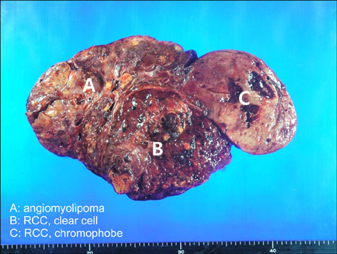
FIG. 2 On gross examination, three different renal masses were found. The cut surface of the largest mass (B) in the midportion had a heterogeneous appearance, composed of hemorrhagic, necrotic, and focal golden yellow soft areas. The cut surface of the smaller mass (C) showed focal hemorrhagic areas in the diffuse yellowish-grayish soft area. In the remaining renal parenchyma, there were multifocal poorly demarcated yellowish, glistening mass-like lesions (A).

FIG. 3 (A) Renal cell carcinoma (RCC), clear-cell type, composed of nests of cells with clear cytoplasm, surrounded by abundant thin-walled blood vessels. (B) RCC, chromophobe type, composed of nests of cells with abundant cytoplasm, sharply outlined cell membranes, raisinoid nuclei, and perinuclear halos.
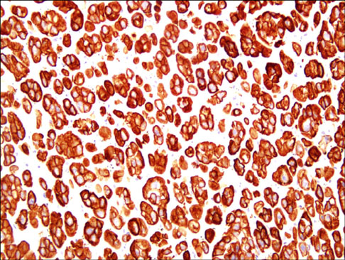
FIG. 4 Renal cell carcinoma (RCC), chromophobe type, showing intense immunoreactivity for cytokeratin 7.
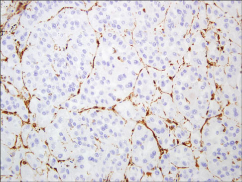
FIG. 5 Renal cell carcinoma (RCC), chromophobe type, showing complete absence of reactivity for vimentin within the tumor cells, except for sinusoidal blood vessels.
Reference
-
1. Störkel S, Eble JN, Adlakha K, Amin M, Blute ML, Bostwick DG, et al. Classification of renal cell carcinoma: Workgroup No. 1. Union Internationale Contre le Cancer (UICC) and the American Joint Committee on Cancer (AJCC). Cancer. 1997. 80:987–989.2. Jimenez RE, Eble JN, Reuter VE, Epstein JI, Folpe AL, de Peralta-Venturina M, et al. Concurrent angiomyolipoma and renal cell neoplasia: a study of 36 cases. Mod Pathol. 2001. 14:157–163.3. Licht MR, Novick AC, Tubbs RR, Klein EA, Levin HS, Streem SB. Renal oncocytoma: clinical and biological correlates. J Urol. 1993. 150:1380–1383.4. Roehrl MH, Selig MK, Nielsen GP, Dal Cin P, Oliva E. A renal cell carcinoma with components of both chromophobe and papillary carcinoma. Virchows Arch. 2007. 450:93–101.5. Morelli L, Pusiol T, Piscioli I, Larosa M, Pozzoli GL, Monica B. Concurrent occurrence of three primary neoplasms with different hystotype in the same kidney, associated with an adenoma of the omolateral adrenal gland: first case report. Int J Urol. 2006. 13:1236–1239.6. Schneck FX, Banner BF, Bahnson RR. Multiple renal neoplasms: a case of 3 histologically dissimilar primary tumors. J Urol. 1991. 145:1251–1253.7. Dincol D, Arican A, Ensari A, Akyar S, Bedük Y, Cengiz A. Concurrent occurrence of three neoplasms including non-Hodgkin's lymphoma, renal cell carcinoma and leiomyoma in the same kidney. Med Oncol. 1999. 16:134–138.8. Tyritzis SI, Alexandrou PT, Migdalis V, Koritsiadis G, Anastasiou I. Synchronous chromophobe and papillary renal cell carcinoma. First report and review of the pathogenesis theories. Pathol Int. 2009. 59:193–196.9. Jun SY, Cho KJ, Kim CS, Ayala AG, Ro JY. Triple synchronous neoplasms in one kidney: report of a case and review of the literature. Ann Diagn Pathol. 2003. 7:374–380.10. Bjornsson J, Short MP, Kwiatkowski DJ, Henske EP. Tuberous sclerosis-associated renal cell carcinoma. Clinical, pathological, and genetic features. Am J Pathol. 1996. 149:1201–1208.
- Full Text Links
-
- Actions
-
Cited
- CITED
-
- Close
- Share
-
- Similar articles
-
- Bilateral Renal Angiomyolipomas without Tuberous Sclerosis
- Bilateral Renal Angiomyolipomas with Tuberous Sclerosis
- Renal and hepatic angiomyolipoma and renal failure in two cases of tuberous sclerosis
- A case of bilateral renal angiomyolipoma associated with tuberous sclerosis
- Anesthetic Management of a Patient with Tuberous Sclerosis for Selective Arterial Embolization: A case report
