Korean J Urol.
2008 Jul;49(7):656-658.
A Large Stone in the Dilated Left Seminal Vesicle: Laparoscopic Removal and Partial Seminal Vesiculectomy
- Affiliations
-
- 1Department of Urology, Chungbuk National University College of Medicine, Cheongju, Korea. wjkim@chungbuk. ac.kr
- 2Department of Urology, School of Medicine, Kyung Hee University, Seoul, Korea.
Abstract
- Stones in the seminal vesicle are extremely rare. We report a case with a large stone in a dilated seminal vesicle. A 20-year-old man presented with a large calcified density in the pelvic cavity on plain films. A 6.0 cm cone shaped stone was noted in the dilated left seminal vesicle diagnosed by radiological examination. We treated the patient by transperitoneal laparoscopic stone removal and partial seminal vesiculectomy. The composition of stone was carbonate apatite. This approach to the treatment of such pathological conditions of the seminal vesicles provides an additional option.
Keyword
Figure
-
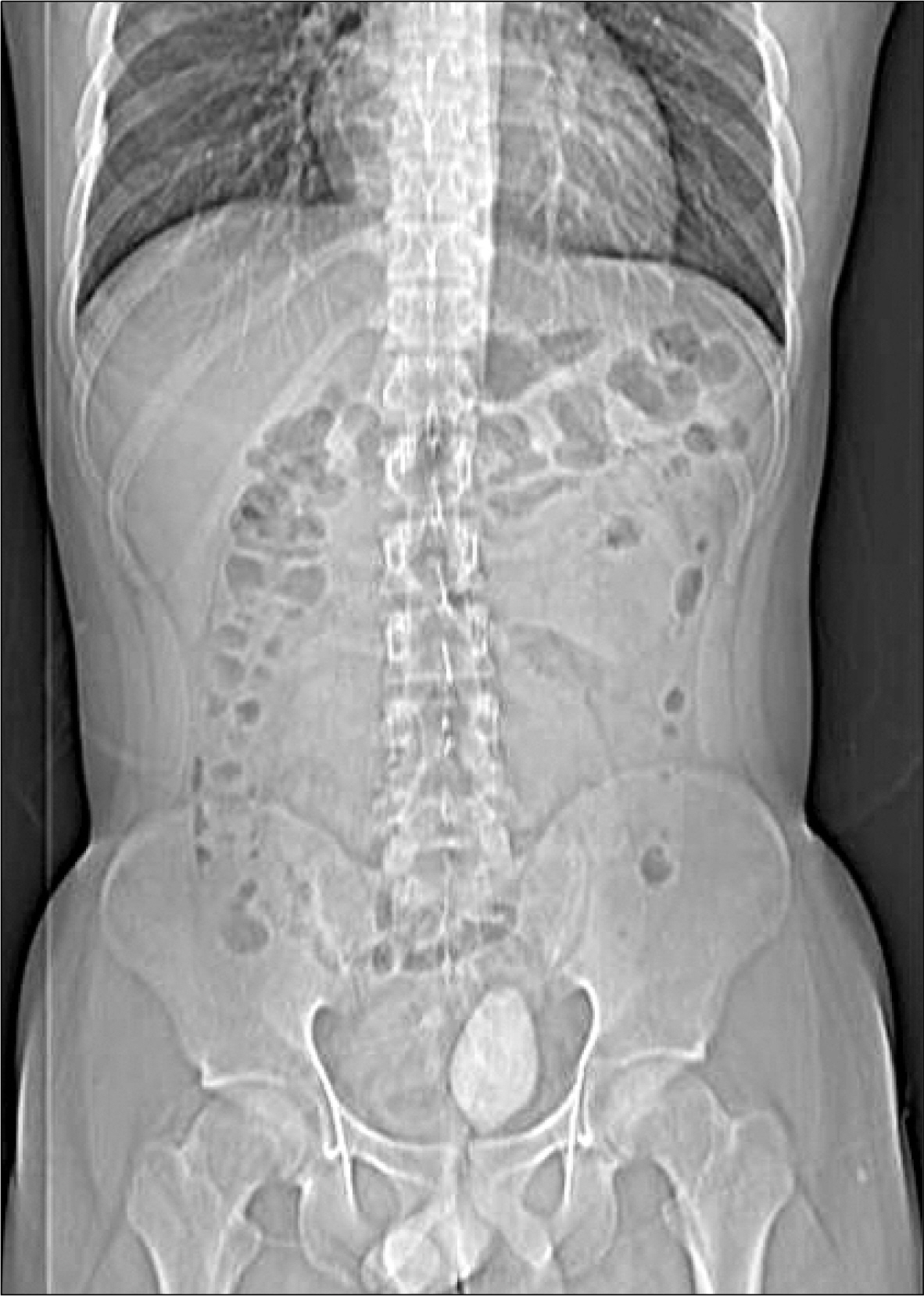
Fig. 1. Plain film shows a large calcified density in the pelvic cavity.

Fig. 2. CT scan demonstrates a large stone in a dilated left seminal vesicle.
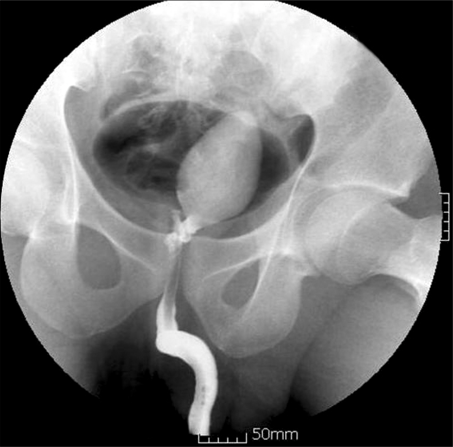
Fig. 3. Retrograde urethrogram reveals reflux into the left seminal vesicle with the stone.
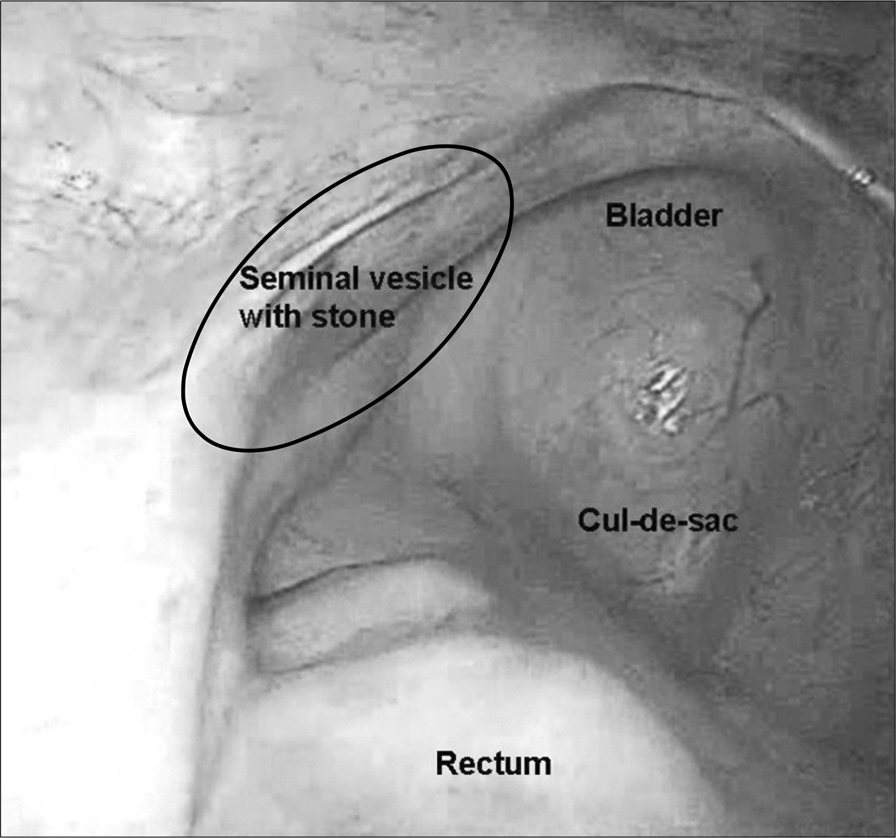
Fig. 4. Laparoscopic view of the cul-de-sec and associated organs.
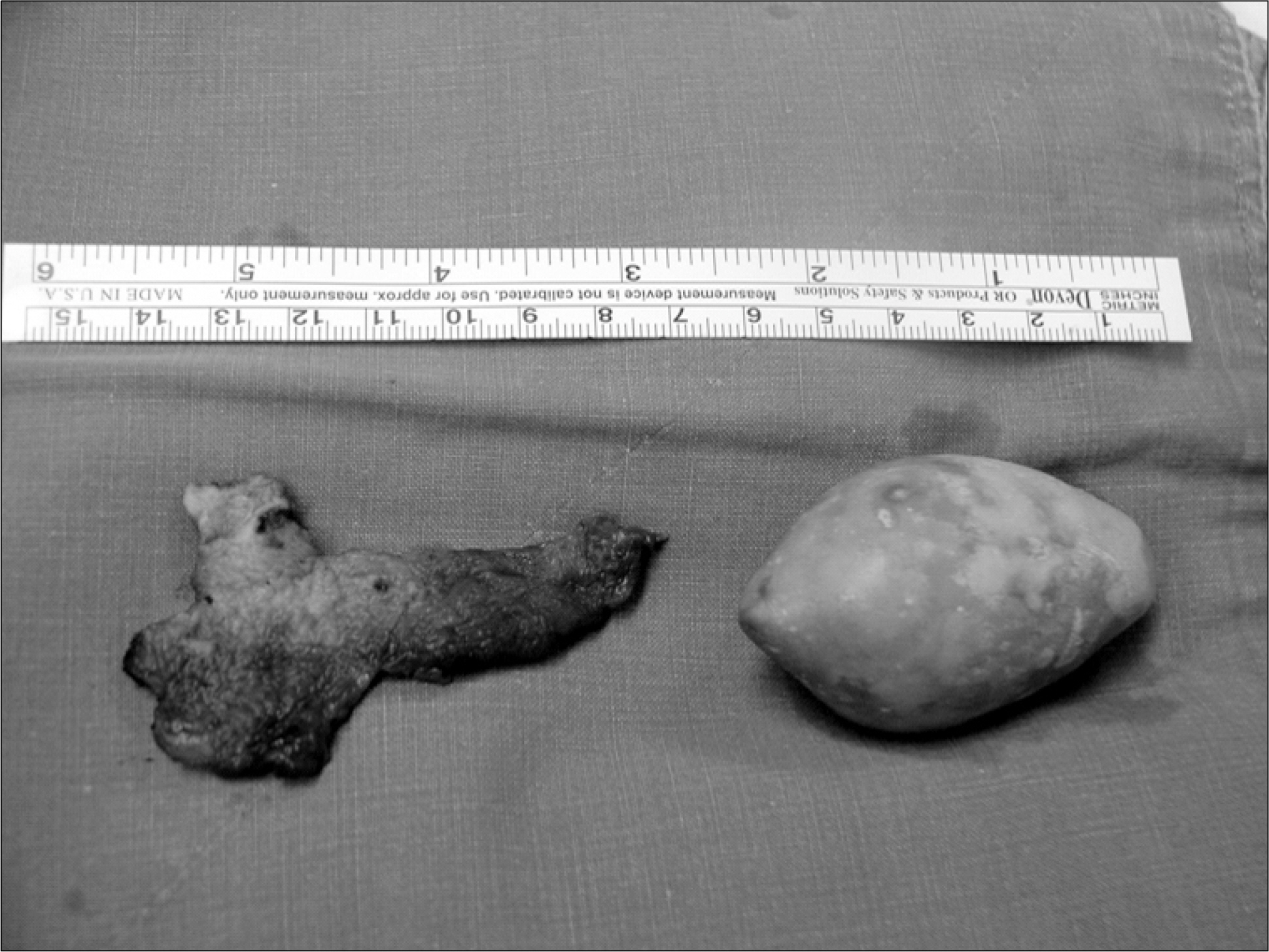
Fig. 5. Removed stone and the dilated left seminal vesicle wall.
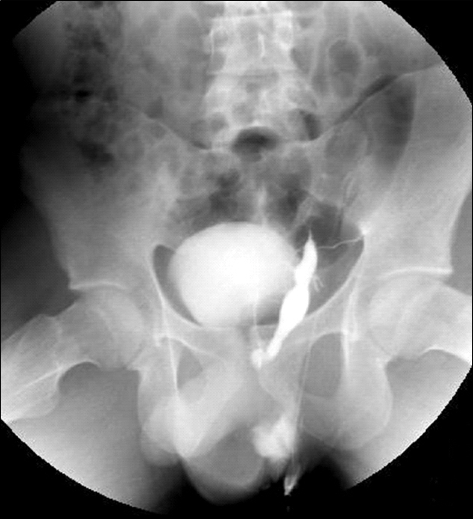
Fig. 6. Postoperative vasogram shows the decreased size of the left seminal vesicle without the stones or leak.
Reference
-
1.Amano T., Kunimi K., Ohkawa M. Transrectal ultrasonography of the prostate and seminal vesicles with hemospermia. Urol Int. 1994. 53:139–42.
Article2.Sandlow JI., Winfield HN., Goldstain M. Surgery of the scrotum and seminal vesicles. Wein AJ, Kavoussi LR, Novick AC, Partin AW, Peters CA, editors. editors.Campbell-Walsh urology. 9th ed.Philadelphia: Saunders;2007. 1098-127.3.Modi PR. Case report: endoscopic management of seminal vesicle stones with cutaneous fistula. J Endourol. 2006. 20:432–5.
Article4.Cuda SP., Brand TC., Thibault GP., Stack RS. Case report: endoscopic laser lithotripsy of seminal-vesicle stones. J Endo-urol. 2006. 20:916–8.
Article5.Uchijima Y., Hiraga S., Akutsu M., Yoshida K., Hobo M., Okada K. Stones of the seminal vesicles and ejaculatory duct in infant: report of a case. Hinyokika Kiyo. 1984. 30:1843–9.6.Carachi R., Gobara D. Recurrent epididymo-orchitis in a child secondary to a stone in the seminal vesicle. Br J Urol. 1997. 79:997.
Article7.Li YK. Diagnosis and management of large seminal vesicle stones. Br J Urol. 1991. 68:322–3.
Article8.Wesson L., Steinhardt G. Case profile: seminal vesicle stones. Urology. 1983. 22:204–5.
Article9.Orquiza CS., Bhayani BN., Berry JL., Dahlen CP. Ectopic opening of the ureter into the seminal vesicle: report of case. J Urol. 1970. 104:532–5.
Article10.Yang SC., Rha KH., Byon SK., Kim JH. Transutricular seminal vesiculoscopy. J Endourol. 2002. 16:343–5.
Article
- Full Text Links
-
- Actions
-
Cited
- CITED
-
- Close
- Share
-
- Similar articles
-
- Laparoscopic Excision of Congenital Seminal Vesicle Cyst Associated with Ipsilateral Renal Agenesis
- Effect of the Vasectomy and Seminal Vesiculectomy on the Fine Structure of the Sperm-Acrosome
- Laparoscopic Excision of Giant Seminal Vesicle Cyst Associated with Ipsilateral Renal Agenesis
- Seminal Vesicle Infection of Zinner Syndrome Misdiagnosed for Neoplasm
- The findings of transrectal ultrasonography in evaluation of organic hemospermia
