Korean J Urol.
2008 Dec;49(12):1125-1130.
Clinical Course of Prenatally-detected Hydronephrosis: Focus on Ureteropelvic Junction Obstruction
- Affiliations
-
- 1Department of Urology, College of Medicine, Gyeongsang National University, Jinju, Korea. kychung@gshp.gsnu.ac.kr
Abstract
-
PURPOSE: The diagnosis and treatment of prenatally-diagnosed hydronephrosis remain controversial. We have conducted a retrospective study to examine the clinical characteristics and course of prenatally-diagnosed hydronephrosis, especially when in the presence of ureteropelvic junction obstruction(UPJO).
MATERIALS AND METHODS
Among all pediatric patients diagnosed with hydronephrosis by prenatal ultrasonography between September 2002 and June 2008, the study was performed on 103 patients(126 renal units), and the mean follow-up period was 19.2 months(range, 6-24 months). Ultrasonography was performed 2-3 days after birth, and after 1, 3, 6, and 12 months, and annually thereafter. Hydronephrosis was graded according to the Society for Fetal Urology(SFU) classification guidelines.
RESULTS
On ultrasonography performed 2-3 days after birth hydronephrosis was graded as follows: grade 1, 45(35.7%); grade 2, 49(38.9%); grade 3, 23(18.3%); and grade 4, 9(7.1%) renal units. In cases with UPJO complete improvement of hydronephrosis was detected in 16 renal units(40%); the renal units and rate of complete improvement in grades 1, 2, 3, and 4 were 12(75%), 3(17.6%), 1(16.7%), and 0(0%), respectively. The anticipated times of complete improvement of hydronephrosis in UPJO grades 1, 2, and 3 were 22.0, 31.3, and 50.4 months, respectively.
CONCLUSIONS
In UPJO, the possibility of improvement of hydronephrosis lower than grade 2 was high, and thus follow-up for approximately 30 months may be needed. In patients with hydronephrosis >grade 3, the rate of improvement was low, thus compulsive follow-up is required.
Keyword
MeSH Terms
Figure
-
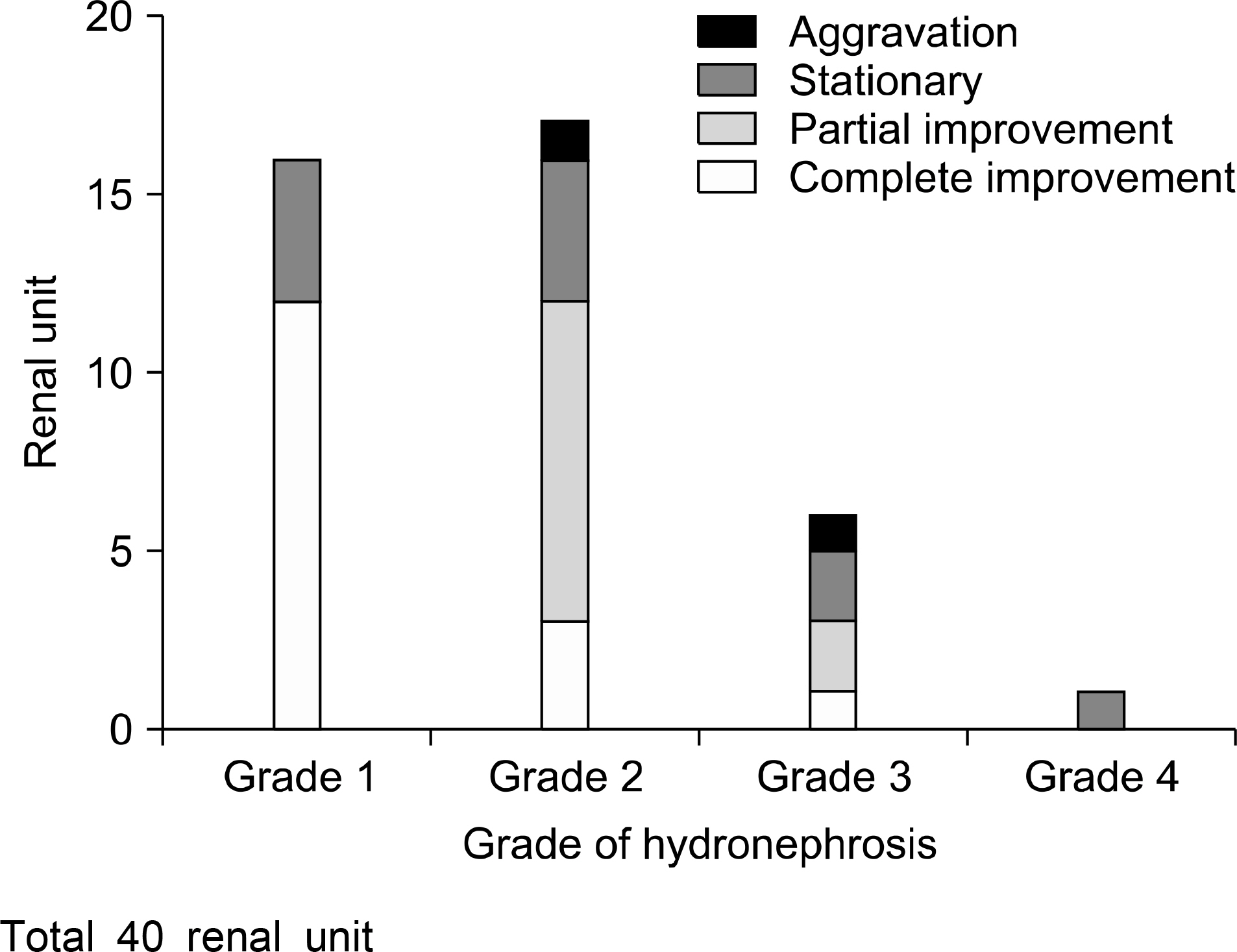
Fig. 1. Clinical course of hydronephrosis in ureteropelvic junction obstruction.
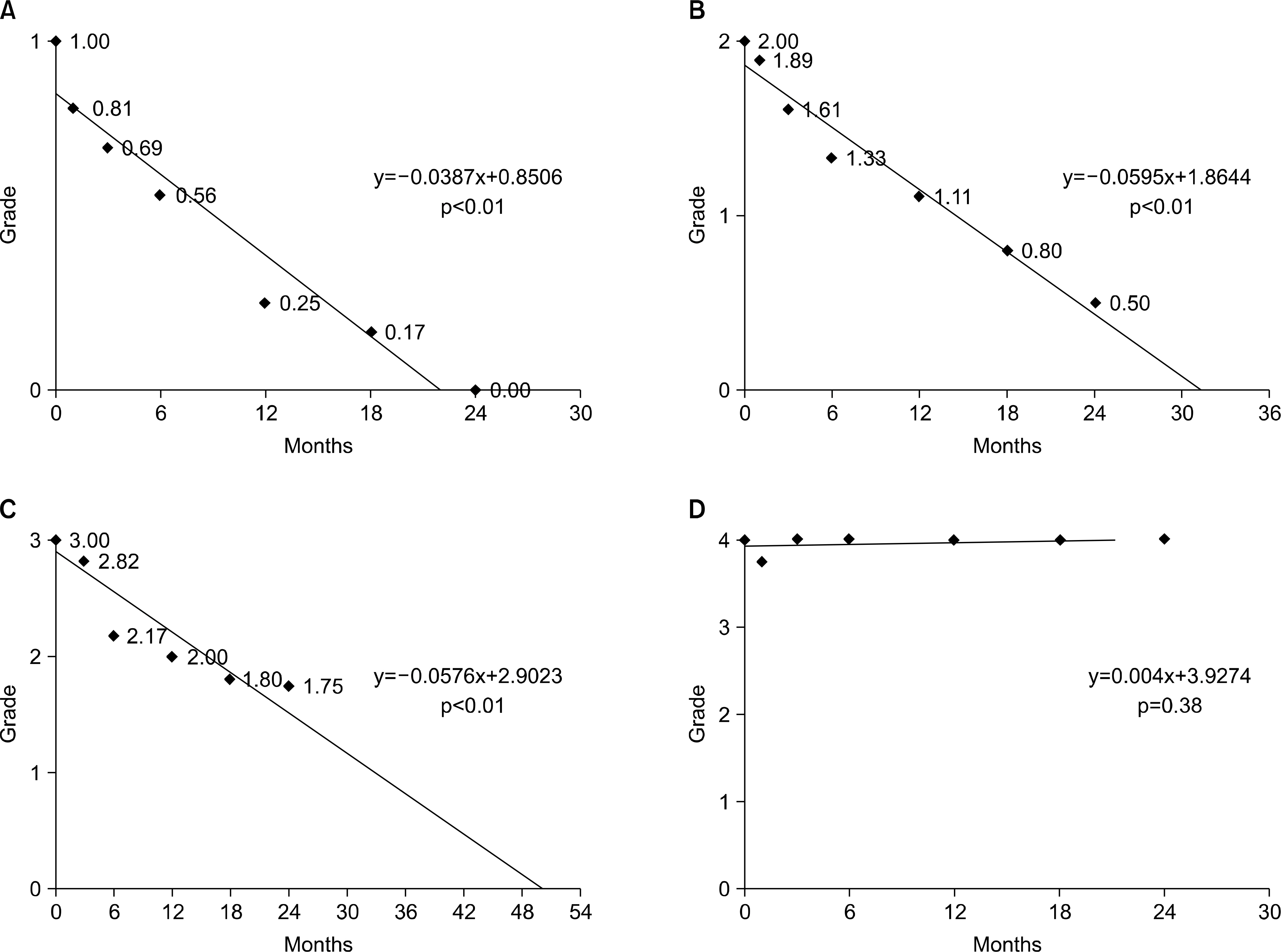
Fig. 2. Expected improvement time of hydronephrosis in ureteropelvic junction obstruction. (A) Grade 1 (total 16 renal units) and linear regression trend line. (B) Grade 2 (total 18 renal units) and linear regression trend line. (C) Grade 3 (total 11 renal units) and linear regression trend line. (D) Grade 4 (total 4 renal units) and linear regression trend line.
Reference
-
References
1. Cho JS, Kim KS, Kim SY, Kim TY, Choi HM, Lim YK, et al. Congenital anomalies diagnosed by antenatal ultrasonography. Korean J Obstet Gynecol. 1997; 40:1228–32.2. O'Flynn KJ, Gough DC, Gupta S, Lewis MA, Postlethwaite RJ. Prediction of recovery in antenatally diagnosed hydronephrosis. Br J Urol. 1993; 71:478–80.3. Benacerraf BR, Mandell J, Estroff JA, Harlow BL, Frigoletto FD Jr. Fetal pyelectasis: a possible association with Down syndrome. Obstet Gynecol. 1990; 76:58–60.4. Fernbach SK, Maizels M, Conway JJ. Ultrasound grading of hydronephrosis: introduction to the system used by the Society for Fetal Urology. Pediatr Radiol. 1993; 23:478–80.
Article5. Roth JA, Diamond DA. Prenatal hydronephrosis. Curr Opin Pediatr. 2001; 13:138–41.
Article6. Helin I, Persson PH. Prenatal diagnosis of urinary tract abnormalities by ultrasound. Pediatrics. 1986; 78:879–83.
Article7. Alladi A, Agarwala S, Gupta AK, Bal CS, Mitra DK, Bhatnagar V. Postnatal outcome and natural history of antenatally-detected hydronephrosis. Pediatr Surg Int. 2000; 16:569–72.
Article8. Ulman I, Jayanthi VR, Koff SA. The longterm followup of newborns with severe unilateral hydronephrosis initially treated nonoperatively. J Urol. 2000; 164:1101–5.
Article9. Hafez AT, McLorie G, Bagli D, Khoury A. Analysis of trends on serial ultrasound for high grade neonatal hydronephrosis. J Urol. 2002; 168:1518–21.
Article10. Lim DJ, Lee HM, Choi H. Clinical characteristics and outcome of fetal hydronephrosis. Korean J Urol. 2004; 45:29–33.11. Ransley PG, Dhillon HK, Gordon I, Duffy PG, Dillon MJ, Barratt TM. The postnatal management of hydronephrosis diagnosed by prenatal ultrasound. J Urol. 1990; 144:584–7.
Article12. Najmaldin AS, Burge DM, Atwell JD. Outcome of antenatally diagnosed pelviureteric junction hydronephrosis. Br J Urol. 1991; 67:96–9.13. Harding LJ, Malone PS, Wellesley DG. Antenatal minimal hydronephrosis: is its follow-up an unnecessary cause of concern? Prenat Diagn. 1999; 19:701–5.
Article14. Park JH, Lee YT, Shin JS. The clinical course of mild neonatal hydronephrosis. Korean J Urol. 2000; 41:872–7.15. Fine RN. Diagnosis and treatment of fetal urinary tract abnormalities. J Pediatr. 1992; 121:333–41.
Article16. Anderson PA, Rickwood AM. Features of primary vesicoureteric reflux detected by prenatal sonography. Br J Urol. 1991; 67:267–71.
Article
- Full Text Links
-
- Actions
-
Cited
- CITED
-
- Close
- Share
-
- Similar articles
-
- A Case of an Ureteropelvic Junction Obstruction Caused by a Crossing Vessel
- Endopyelotomy as a Treatment for Ureteropelvic Junction Obstruction: 3 Cases
- Urinary Ascites with Urinoma Secondary to Ureteropelvic Junction Obstruction in an Infant: A Case Report
- A Case of Congenital Ureteropelvic Junction Obstruction with more than 500 Stones
- Experiences on Anderson-Hynes Pyeloplasty
