Range of Motion of the Ankle According to Pushing Force, Gender and Knee Position
- Affiliations
-
- 1Department of Rehabilitation Medicine, Chungnam National University Hospital, Daejeon, Korea. asacara@naver.com
- KMID: 2309926
- DOI: http://doi.org/10.5535/arm.2016.40.2.271
Abstract
OBJECTIVE
To investigate the difference of range of motion (ROM) of ankle according to pushing force, gender and knee position.
METHODS
One hundred and twenty-eight healthy adults (55 men, 73 women) between the ages of 20 and 51, were included in the study. One examiner measured the passive range of motion (PROM) of ankle by Dualer IQ Inclinometers and Commander Muscle Testing. ROM of ankle dorsiflexion (DF) and plantarflexion (PF) according to change of pushing force and knee position were measured at prone position.
RESULTS
There was significant correlation between ROM and pushing force, the more pushing force leads the more ROM at ankle DF and ankle PF. Knee flexion of 90° position showed low PF angle and high ankle DF angle, as compared to the at neutral position of knee joint. ROM of ankle DF for female was greater than for male, with no significant difference. ROM of ankle PF for female was greater than male regardless of the pushing force.
CONCLUSION
To our knowledge, this is the first study to assess the relationship between pushing force and ROM of ankle joint. There was significant correlation between ROM of ankle and pushing force. ROM of ankle PF for female estimated greater than male regardless of the pushing force and the number of measurement. The ROM of the ankle is measured differently according to the knee joint position. Pushing force, gender and knee joint position are required to be considered when measuring the ROM of ankle joint.
MeSH Terms
Figure
-
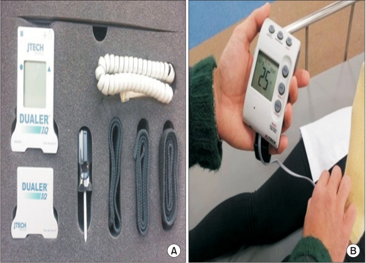
Fig. 1 (A) Dualer IQ Inclinometers (J-Tech, Torrance, CA, USA), (B) Commander Muscle Tester (J-Tech).
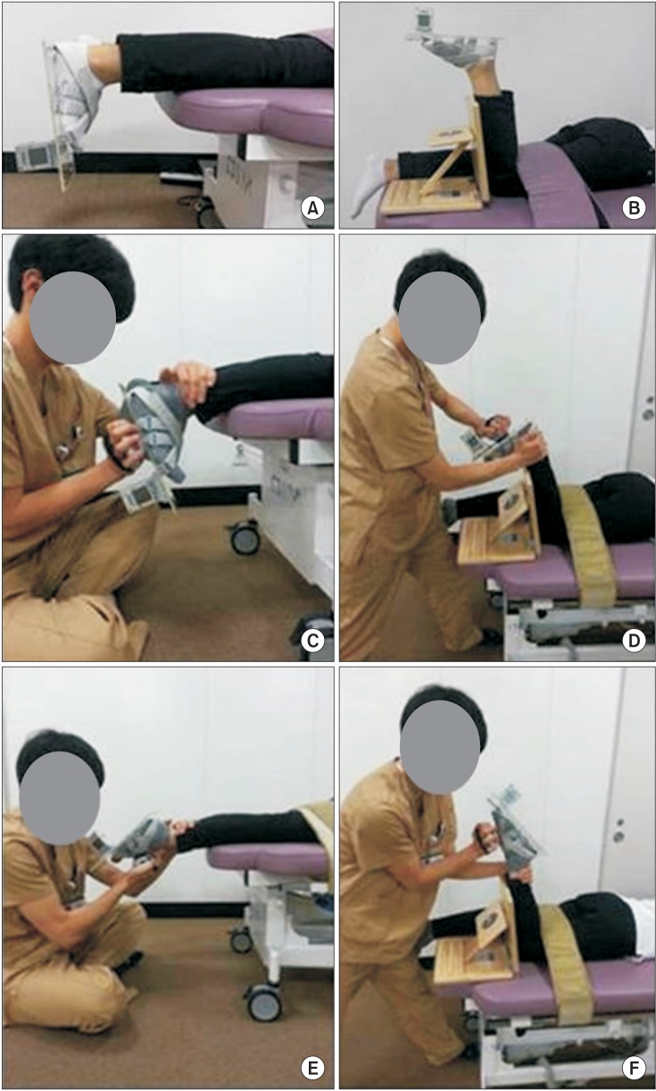
Fig. 2 Starting position before measurement of range of motion of ankle dorsiflexion & plantarflexion at neutral position of knee joint (A) and ankle dorsiflexion & plantarflexion at knee flexion of 90° (B). Position to measurement of range of motion of ankle dorsiflexion (C, D) and ankle plantarflexion (E, F).
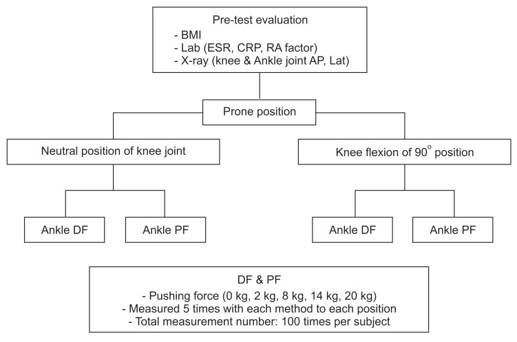
Fig. 3 Overall flow sheet of the measurement process. BMI, body mass index; ESR, erythrocyte sedimentation rate; CRP, C-reactive protein; RA, rheumatoid arthritis; AP, anteroposterior; DF, dorsiflexion; PF, plantarflexion.
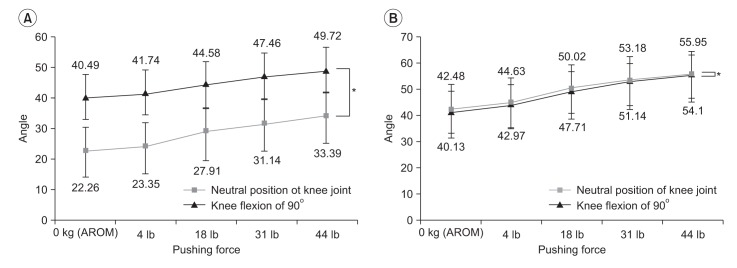
Fig. 4 Measured range of motion (ROM) values of ankle dorsiflexion at neutral position of knee joint and knee flexion of 90° (A) and plantarflexion at neutral position of knee joint and knee flexion of 90° (B) associated with pushing force.

Fig. 5 Differences in the male and female of range of motion (ROM) of ankle dorsiflexion at neutral position of knee joint (A) and at knee flexion of 90° (B).
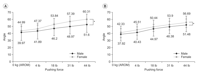
Fig. 6 Differences in the male and female of range of motion (ROM) of ankle plantarflexion at neutral position of knee joint (A) and at knee flexion of 90° (B).
Cited by 1 articles
-
Determining the Reliability of a New Method for Measuring Joint Range of Motion Through a Randomized Controlled Trial
So Young Ahn, Hanbit Ko, Jeong Oh Yoon, Sun Ung Cho, Jong Hyun Park, Kang Hee Cho
Ann Rehabil Med. 2019;43(6):707-719. doi: 10.5535/arm.2019.43.6.707.
Reference
-
1. Wong Y, Kim W, Ying N. Passive motion characteristics of the talocrural and the subtalar joint by dual Euler angles. J Biomech. 2005; 38:2480–2485. PMID: 16214496.
Article2. Leardini A, O'Connor JJ, Catani F, Giannini S. Kinematics of the human ankle complex in passive flexion; a single degree of freedom system. J Biomech. 1999; 32:111–118. PMID: 10052915.
Article3. Roaas A, Andersson GB. Normal range of motion of the hip, knee and ankle joints in male subjects, 30-40 years of age. Acta Orthop Scand. 1982; 53:205–208. PMID: 7136564.
Article4. American Academy of Orthopaedic Surgeons. Joint motion: methods of measuring and recording. Edinburgh: Churchill Livingstone;1966.5. Ahlberg A, Moussa M, Al-Nahdi M. On geographical variations in the normal range of joint motion. Clin Orthop Relat Res. 1988; 234:229–231. PMID: 3409581.
Article6. James B, Parker AW. Active and passive mobility of lower limb joints in elderly men and women. Am J Phys Med Rehabil. 1989; 68:162–167. PMID: 2765206.
Article7. Macedo LG, Magee DJ. Differences in range of motion between dominant and nondominant sides of upper and lower extremities. J Manipulative Physiol Ther. 2008; 31:577–582. PMID: 18984240.
Article8. Youdas JW, Bogard CL, Suman VJ. Reliability of goniometric measurements and visual estimates of ankle joint active range of motion obtained in a clinical setting. Arch Phys Med Rehabil. 1993; 74:1113–1118. PMID: 8215866.
Article9. Grimston SK, Nigg BM, Hanley DA, Engsberg JR. Differences in ankle joint complex range of motion as a function of age. Foot Ankle. 1993; 14:215–222. PMID: 8359768.
Article10. Kumar S, Sharma R, Gulati D, Dhammi IK, Aggarwal AN. Normal range of motion of hip and ankle in Indian population. Acta Orthop Traumatol Turc. 2011; 45:421–424. PMID: 22245818.
Article11. Venturini C, Ituassu NT, Teixeira LM, Deus CV. Intrarater and interrater reliability of two methods for measuring the active range of motion for ankle dorsiflexion in healthy subjects. Rev Bras Fisioter. 2006; 10:407–411.12. de Winter AF, Heemskerk MA, Terwee CB, Jans MP, Deville W, van Schaardenburg DJ, et al. Inter-observer reproducibility of measurements of range of motion in patients with shoulder pain using a digital inclinometer. BMC Musculoskelet Disord. 2004; 5:18. PMID: 15196309.
Article13. Belcjak CE, Cavalheri G, Pereira JM, Caffaro RA. The differences in the ankle range of motion in distinct ethnical groups measured by goniometry. Phlebologie. 2009; 38:59–63.
Article14. Keating JL, Parks C, Mackenzie M. Measurements of ankle dorsiflexion in stroke subjects obtained using standardised dorsiflexion force. Aust J Physiother. 2000; 46:203–213. PMID: 11676804.
Article15. Moseley A, Adams R. Measurement of passive ankle dorsiflexion: procedure and reliability. Aust J Physiother. 1991; 37:175–181. PMID: 25026482.
Article16. Hallaceli H, Uruc V, Uysal HH, Ozden R, Hallaceli C, Soyuer F, et al. Normal hip, knee and ankle range of motion in the Turkish population. Acta Orthop Traumatol Turc. 2014; 48:37–42. PMID: 24643098.
Article17. Kendall FP, McCreary EK, Provance PG, Rodger MM, Romani WA. Muscle: testing and function with posture and pain. 5th ed. Philadelphia: Saunders;2005. p. 359–464.18. Mitchell B, Bressel E, McNair PJ, Bressel ME. Effect of pelvic, hip, and knee position on ankle joint range of motion. Phys Ther Sport. 2008; 9:202–208. PMID: 19083721.
Article19. Gerlach UJ, Lierse W. Functional construction of the superficial and deep fascia system of the lower limb in man. Acta Anat (Basel). 1990; 139:11–25. PMID: 2288185.
Article20. Cho SH, Park JM, Kwon OY. Gender differences in three dimensional gait analysis data from 98 healthy Korean adults. Clin Biomech (Bristol, Avon). 2004; 19:145–152.
Article21. Chung MJ, Wang MJ. The effect of age and gender on joint range of motion of worker population in Taiwan. Int J Ind Ergon. 2009; 39:596–600.
Article22. Baldwin JN, McKay MM, Hiller CE, Nightingale JE, Moloney N, Vanicek N, et al. 1000 Norms Project: understanding foot and ankle health, disease and normality. J Foot Ankle Res. 2014; 7(Suppl 1):A6.
Article
- Full Text Links
-
- Actions
-
Cited
- CITED
-
- Close
- Share
-
- Similar articles
-
- Changes of Length and Active Force of the Soleus Muscle According to the Position of Ankle Joint in Rabbit
- The Effects of Ankle Plantar Flexors Stretching Exercise on Functional Reach in Elderly Men
- Does contralateral knee range of motion predict postoperative knee range of motion after total knee arthroplasty?
- Combined Effect of Vibration Intensity, Grip Temperature, Noise and Pushing Power on Grip Forces and Skin Temperatures of Fingers
- The Effects of Changes of Ankle Strength and Range of Motion According to Aging on Balance
