A Case of Infected Left Atrial Myxoma With Concomitant Mitral Valve Endocarditis
- Affiliations
-
- 1Division of Cardiology, Department of Internal Medicine, Samyook Medical Center, Seoul, Korea. mulgang@gmail.com
- 2Division of Infection, Department of Internal Medicine, Samyook Medical Center, Seoul, Korea.
- 3Department of Cardiac Surgery, Samyook Medical Center, Seoul, Korea.
- 4Department of Pathology, Samyook Medical Center, Seoul, Korea.
- KMID: 2297923
- DOI: http://doi.org/10.4070/kcj.2011.41.10.618
Abstract
- Myxoma is the most common primary tumor in the heart. Cardiac myxomas can present in various manners including embolization and fever, sometimes simulating endocarditis. However, they are rarely infected. We report here a case of an infected left atrial myxoma that seeded a normal mitral valve and atypically presented with multiple embolic events in the lower extremities along with multiple splenic and a cerebellar infarction.
Keyword
Figure
-
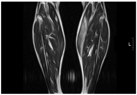
Fig. 1 T2-weighted image of the left gastrocnemius. This image shows an abnormally ill defined high signal that suggests inflammation in the medial belly of the left gastrocnemius.
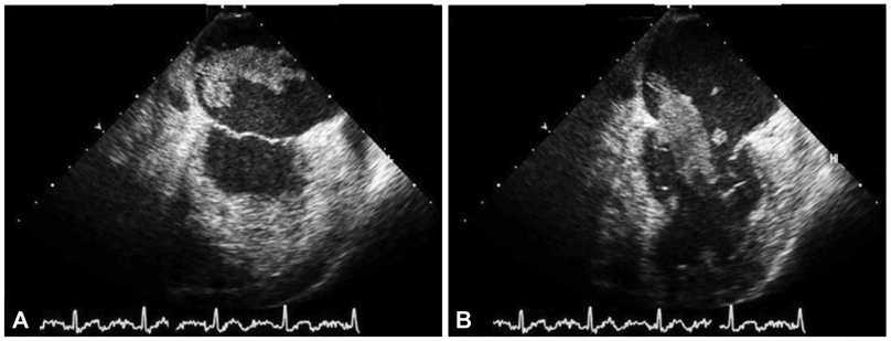
Fig. 2 Transesophageal echocardiography. A: echocardiography shows a left atrial tumor of 5.6×1.8 cm with a villous surface in the systole. B: the mass protrudes from the left atrium into the left ventricle in the diastole.
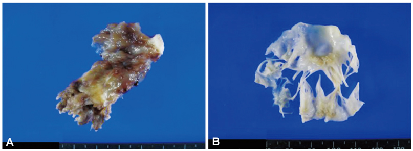
Fig. 3 Gross specimen of the mass and the mitral valve. A: the mass has a villous surface with a base of about 9 mm. B: multiple tiny and nodular vegetations of less than 1 mm are found on the P2 and P3 portion of the posterior mitral leaflet.
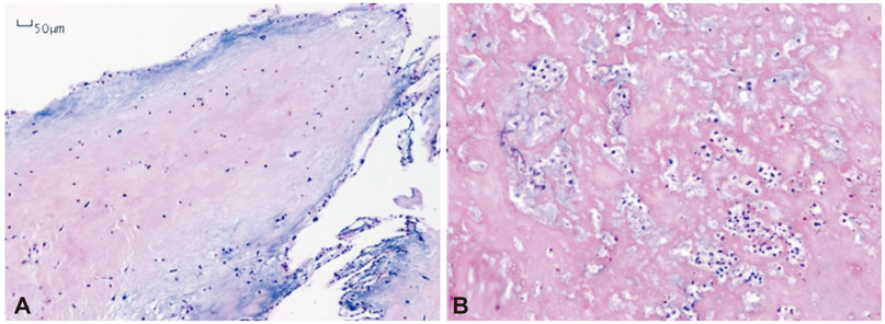
Fig. 4 Microscopic features of the specimen. A: the mass is composed of small round polygonal cells in the loose myxoid stroma (Hematoxylin and Eosin staining, 40× magnification). B: higher magnification of the superficial lesion reveals aggregates of polymorphonuclear neutrophils in degenerating stroma, suggesting microabscess (Hematoxylin and Eosin staining, 200× magnification).
Reference
-
1. Rhim HY, Youn HJ, Park JW, et al. Clinical experience of cardiac myxoma. Korean Circ J. 1999. 29:1317–1323.2. Kim CJ, Doh MH, Kwon OH, et al. Clinical observation of cardiac myxoma. Korean Circ J. 1985. 15:671–679.3. Llinares P, Mlguue E, Echamz A, Juffé A, Prado G, Alvarez N. Infect-ed atrial myxoma simulating infective endocarditis. Enferm Infecc Microbiol Clin. 1993. 11:378–381.4. Whitman MS, Rovito MA, Klions D, Tunkel AR. Infected atrial myxoma: case report and review. Clin Infect Dis. 1994. 18:657–658.5. Uchino K, Mochida Y, Ebina T, et al. Infected left atrial myxoma. Intern Med. 2002. 41:957–960.6. Tunick PA, Fox AC, Culliford A, Levy R, Kronzon I. The echocardiographic recognition of an atrial myxoma vegetation. Am Heart J. 1990. 119:679–680.7. Law A, Honos G, Huynh T. Negative predictive value of multiplane transesophageal echocardiography in the diagnosis of infective endocarditis. Eur J Echocardiogr. 2004. 5:416–421.8. Duval X, Papastamopoulos V, Longuet P, et al. Definite Streptococcus bovis endocarditis: characteristics in 20 patients. Clin Microbiol Infect. 2001. 7:3–10.9. Klein RS, Recco RA, Catalano MT, Edberg SC, Casey JI, Stigbigel NH. Association of Streptococcus bovis with carcinoma of the colon. N Engl J Med. 1977. 297:800–802.10. Dekkers P, Elbers HR, Morshuis WJ, Jaarsma W. Infected left atrial myxoma. J Am Soc Echocardiogr. 2001. 14:644–645.11. Kim H, Kang JK, Chung YS, Kim YH, Chung WS. Infected left atrial myxoma. Korean J Thorac Cardiovasc Surg. 2009. 42:513–515.12. Eishi K, Kawazoe K, Kuriyama Y, Kitoh Y, Kawashima Y, Omae T. Surgical management of infective endocarditis associated with cerebral complications. Multi-center retrospective study in Japan. J Thorac Cardiovasc Surg. 1995. 110:1745–1755.
- Full Text Links
-
- Actions
-
Cited
- CITED
-
- Close
- Share
-
- Similar articles
-
- Left Atrial Mural Endocarditis Diagnosed by Transesophageal Echocardiography in a Patient with Mitral Valve Prolapse
- Pregnancy, Paroxysmal Nocturnal Dyspnea, Presyncope, and Plop: A Case of Left Atrial Myxoma Causing Mitral Valve Obstruction in Pregnancy
- Infective Left Atrial Dissecting Flap after Cardiac Surgery
- Left Atrial Dissection by Aorto-Left Atrial Fistula after Aortic Valve Replacement: A case report
- Cardiac Myxoma Originating from the Anterior Mitral Valve Leaflet
