Primary Fibrosarcoma of the Breast: A Case Report
- Affiliations
-
- 1Department of Radiology, Dongguk University Ilsan Hospital, Dongguk University College of Medicine, Goyang, Korea. dbkim@dumc.or.kr
- 2Department of Surgery, Dongguk University Ilsan Hospital, Dongguk University College of Medicine, Goyang, Korea.
- 3Department of Pathology, Dongguk University Ilsan Hospital, Dongguk University College of Medicine, Goyang, Korea.
- KMID: 2286517
- DOI: http://doi.org/10.4048/jbc.2011.14.2.156
Abstract
- A primary fibrosarcoma of the breast is a rare tumor. Here we report on a case of a primary fibrosarcoma of the breast that presented as a palpable left breast mass in a 47-year-old woman. The physical examination revealed a 3 cm sized, round mass in the left upper outer breast. The mammograms revealed a 3 cm sized, partially circumscribed and partially obscured, high density mass in the upper outer quadrant of the left breast. An ultrasonogram demonstrated a 3 cm sized, ovoid, circumscribed and hypoechoic mass with peripheral increased vascularity on Doppler imaging. Surgical excision was performed and the pathology revealed a low grade fibrosarcoma.
Keyword
Figure
-
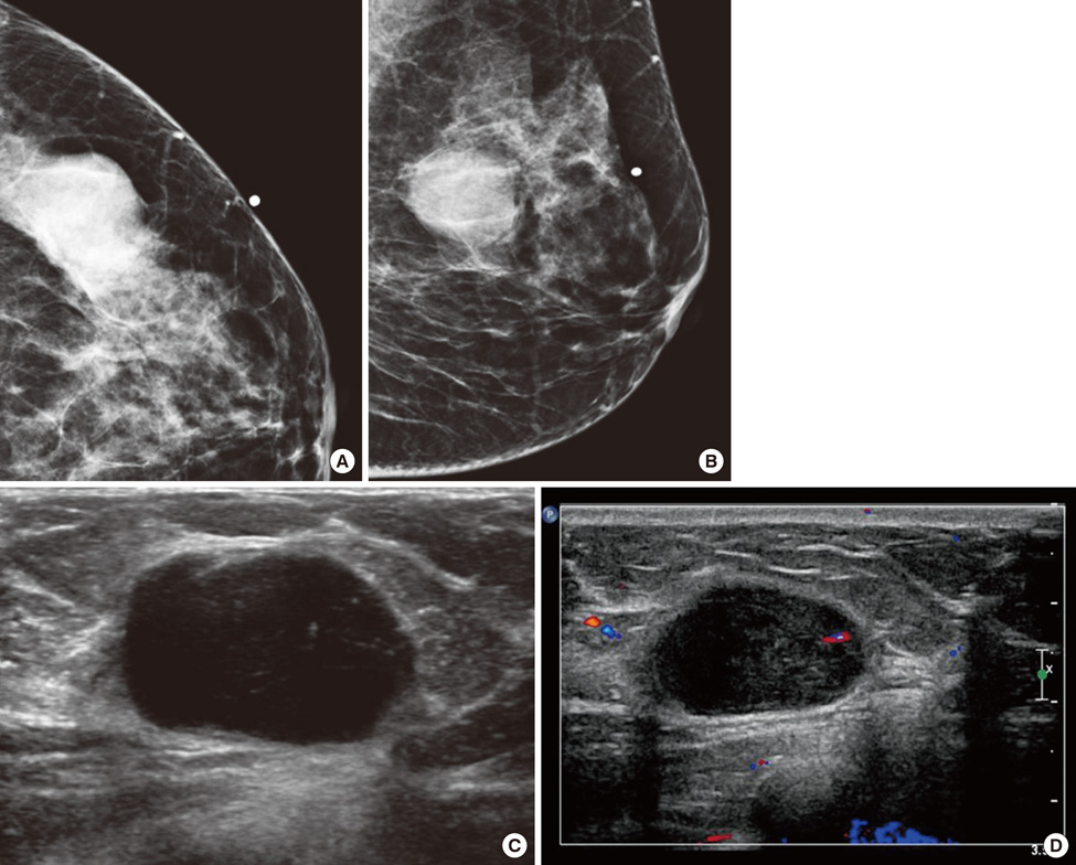
Figure 1 Imaging findings. (A, B) 47-year-old woman resented with a palpable mass in the left breast. Cranicaudal (A) and mediolateral oblique views (B) of a mammogram show an ovoid, partially circumscribed and partially obscured, high-density mass in the upper outer quadrant of the left breast. (C) An ultrasonogram shows an ovoid circumscribed, and homogenous hypoechoic mass with posterior acoustic enhancement in the left breast. (D) The Doppler ultrasonogram shows vascularity in the peripheral portion of the mass.
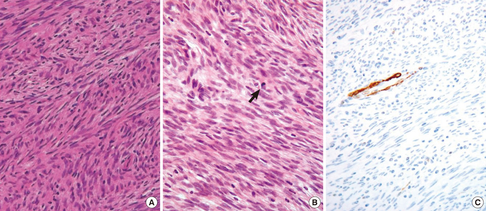
Figure 2 Microscopic findings. (A) Microscopic findings shows spindle cell tumor that has a cell pattern of columns of short parallel lines with all the lines in one column sloping one way and lines in adjacent columns sloping the other way (herring bone pattern) (H&E stain, ×400). (B) Microscopic findings show celluar spindle cells with elongated nuclei and mild to moderate cytologic atypia arranged in interwoven bundles and intermittent mitotic figures (arrow) (H&E stain, ×400). (C) Immunohistochemical finding shows negative reaction to smooth muscle actin immunostain on spindle tumor cells (smooth muscle actin Immunostain, ×400).
Reference
-
1. Barnes L, Pietruszka M. Sarcomas of the breast: a clinicopathologic analysis of ten cases. Cancer. 1977. 40:1577–1585.
Article2. Roberson GV. Fibrosarcoma of the breast. J Ark Med Soc. 1973. 69:257–265.3. Adem C, Reynolds C, Ingle JN, Nascimento AG. Primary breast sarcoma: clinicopathologic series from the Mayo Clinic and review of the literature. Br J Cancer. 2004. 91:237–241.
Article4. Fletcher CD, Unni KK, Mertens F. World Health Organization Classification of Tumours: Pathology and Genetics of Tumours of Soft Tissue and Bone. 2002. Lyon: IARC Press.5. Pollard SG, Marks PV, Temple LN, Thompson HH. Breast sarcoma. A clinicopathologic review of 25 cases. Cancer. 1990. 66:941–944.
Article6. Trent II JC 2nd, Benjamin RS, Valero V. Primary Soft Tissue Sarcoma of the Breast. Curr Treat Options Oncol. 2001. 2:169–176. McDivitt R, Stewart FW, Berg JW. Tumours of the breast. In: Atlas of Tumor Pathology. Washington, D.C.: Armed Forces Institute of Pathology; 1968. p.127-30.
Article7. Hefny AF, Bashir MO, Joshi S, Branicki FJ, Abu-Zidan FM. Stromal sarcoma of the breast: a case report. Asian J Surg. 2004. 27:339–341. Moore MP, Kinne DW. Breast sarcoma. Surg Clin North Am 1996;76: 383-92.
Article8. Terrier P, Terrier-Lacombe MJ, Mouriesse H, Friedman S, Spielmann M, Contesso G. Primary breast sarcoma: a review of 33 cases with immunohistochemistry and prognostic factors. Breast Cancer Res Treat. 1989. 13:39–48.
Article9. Blanchard DK, Reynolds CA, Grant CS, Donohue JH. Primary nonphylloides breast sarcomas. Am J Surg. 2003. 186:359–361.
Article10. Elson BC, Ikeda DM, Andersson I, Wattsgård C. Fibrosarcoma of the breast: mammographic findings in five cases. AJR Am J Roentgenol. 1992. 158:993–995.
Article11. Edge SB, Byrd DR, Compton CC, Fritz AG, Greene FL, Trotti A. AJCC Cancer Staging Manual. 2010. 7th ed. New York: Springer.12. Clinical practice guidelines in oncology - v.2.2010: soft tissue sarcoma. National Comprehensive Cancer Network. Accessed June 23rd, 2010. http://www.nccn.org/professionals/physician_gls/f_guidelines.asp.
